
- Title
-
Zebrafish sip1a and sip1b are essential for normal axial and neural patterning
- Authors
- Delalande, J.M., Guyote, M.E., Smith, C.M., and Shepherd, I.T.
- Source
- Full text @ Dev. Dyn.

RT-PCR and in situ analysis of the spatial and temporal expression pattern of zebrafish sip1a and sip1b. A: RT-PCR of zebrafish sip1a and sip1b with primers flaking Exon9 using mRNA isolated from wild type embryos at 0, 24, 48, and 72hpf. Each gene displayed two bands corresponding to two alternatively spliced mRNAs. B-U: Wholemount in situ hybridized embryos hybridized with either a sip1a (B, C, F, G, J, K, N, O, R, S) or sip1b (D, E, H, I, L, M, P, Q, T, U) antisense probe at the indicated developmental stages. The first and third columns are lateral views; the second and fourth columns are dorsal views. Anterior is to the left. Red arrowhead, pre-otic neural crest (G); green arrowhead, post-otic neural crest (G); pink arrowheads, VII/VIII cranial ganglia (H, I, L, M); light blue arrowheads, V cranial ganglia (L, M); black arrowheads, pharyngeal arches (J, N); white arrowheads, gut mesendoderm (N). |

Effect of sip1a and sip1b splice blocking antisense morpholino oligonucleotide injection on survival and morphological development at 24-72hpf. A: Bar graph showing the percent of surviving sip1a SBMO morphants, sip1b SBMO morphants, and sip1a sip1b double morphants at 24, 48, and 72 hpf. The numbers represent the percent of injected embryos surviving at each specific time point ± s.e.m. based on 3 independent experiments. B-M: Lateral views of control (B, F, J), sip1a morphant (C, G, K), sip1b morphant (D, H, L), and sip1a sip1b double morphant (E, I, M) embryos at 24hpf (B-E), 48hpf (F-I), and 72 (J-M) hpf. Anterior is to the left. PHENOTYPE:
|
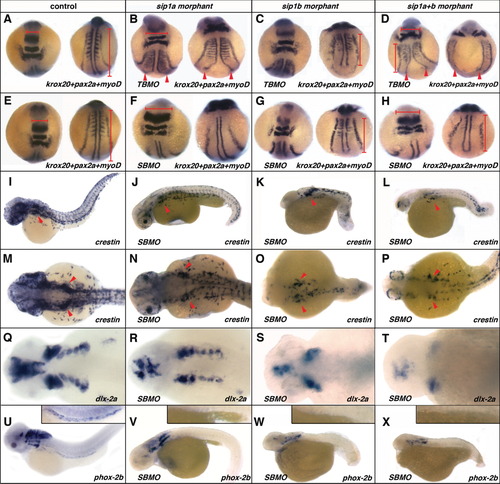
Effect of sip1a and sip1b translation and splice blocking morpholino antisense oligonucleotide injection on axial patterning, and neural crest specification and migration. (A, E, I, M, Q, U) Wild-type control, (B) sip1a TBMO morphant, (F, J, N, R, V) sip1a SBMO morphant, (C) sip1b TBMO morphant, (G, K, O, S) sip1b SBMO morphant embryos, (D, H, L, P, T) sip1a sip1b double morphant embryos. A-H: Dorsal views of 3-somite stage embryos that have been hybridized with a krox20, pax2a, and myoD riboprobes mix. I-L: Lateral views of 36hpf embryos that have been hybridized with riboprobes for crestin. Lateral (M-P) and dorsal (Q-T) views of 48hpf embryos that have been hybridized with riboprobes for dlx-2a. U-X: Lateral views of 55hpf embryos that have been hybridized with riboprobes for phox2b. Insets in U-X are close-ups of the intestine of these embryos. Red arrowheads (B-D) point to convergence defects in the midline. Red horizontal bars (A, B, D, E, F, H) highlight flattening of the morphant embryos in the hindbrain region, as compared to the control. Red vertical bars (A, C, D, E, G, H) highlight shorter axis in morphant embryos, as compared to control. Red arrowheads (I-P) indicate the vagal/post-otic neural crest. Anterior is to the left. EXPRESSION / LABELING:
PHENOTYPE:
|

Unillustrated author statements PHENOTYPE:
|