
- Title
-
Retinoic acid regulation of the Mesp-Ripply feedback loop during vertebrate segmental patterning
- Authors
- Moreno, T.A., Jappelli, R., Izpisúa Belmonte, J.C., and Kintner, C.
- Source
- Full text @ Dev. Biol.

Zebrafish Mesp genes respond differently to RA treatment. (A–D) Zebrafish embryos treated with carrier control (DMSO; top panels) or with RA (lower panels) for 3 h and then stained for the expression of the indicated gene: A, Dr-mespa; B, Dr-mespb; C, mespo; D, mkp3. (E) Zebrafish embryos transgenic for Xl-mespb-GFP were stained for GFP RNA (i), Dr-mespb RNA (ii) or Dr-mespa RNA expression (iii) and aligned according to the first somite. (F) Double-label in situs on zebrafish embryos transgenic for Xl-mespb-GFP with GFP (red) and the gene indicated (purple) to localize the GFP expression pattern. (G) X. laevis (left panel) and zebrafish embryos (right panel) transgenic for the full-length Xl-mespb-GFP were imaged by confocal microscopy. Shown is a region containing newly formed somites, and where somitic boundaries are marked with arrowheads. (H) Zebrafish embryos transgenic for Xl-mespb-GFP were treated with RA at the 6 somite stage (i, ii) or 12 somite stage (iii, iv) and stained for GFP RNA (red) and for no tail (ntl) in purple to mark the notochord. Brackets mark the tailbud domain. Anterior is oriented up in all panels. EXPRESSION / LABELING:
|

Ripply genes modulate the RA response of Mesp genes. (A) Zebrafish embryos treated for 2 h with RA and/or CHX as indicated and stained for the expression Ripply1 (top row) or Ripply2 (bottom row). Anterior is oriented up for zebrafish embryos. (B) Xenopus embryos treated for 1.5 h with RA and/or CHX as indicated and stained for the expression of bowline (top row) or ledgerline (bottom row). Anterior is oriented to the left for frog embryos. (C–E) Zebrafish transgenic for Xl-mespb-GFP embryos were injected with the indicated morpholino: R1 = anti-Ripply1; R2 = anti-Ripply2; R1 + 2 = both morpholinos. Injected embryos were treated for 4 h with DMSO (carrier control) or with RA and then stained for expression of C: mespb; D, mespa; or E, GFP. Anterior is up in all zebrafish panels. (F–I) Xenopus embryos injected with bowlineth RA and then stained for expression of C: mespb; D, mespa; or E, GFP. Anterior is up i (bln, F), ledgerline (ldg, G), both bln and ldg (H), or lacZ RNAs (I). Embryos were treated with DMSO or RA (top and bottom, respectively) and stained for localization of Xl-mespb transcripts. Within each boxed area, the two sides of a single embryo are shown. The uninjected side serves as an internal control and is always shown on the left side of the box. EXPRESSION / LABELING:
|

(A) Live image of an Xl-mespb-GFP transgenic embryo at approximately 20 hpf. (B) GFP fluorescence in an Xl-mespb-GFP transgenic embryo, with strong expression seen in the anterior portion of newly formed somites (arrowhead). EXPRESSION / LABELING:
|
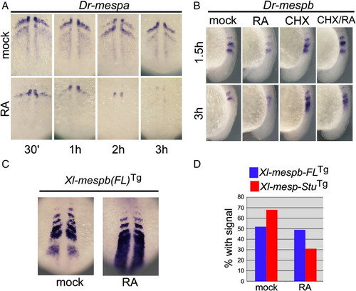
(A) Zebrafish embryos either mock-treated or treated with RA for in the indicated times and then fixed and stained for Dr-mespa RNA. Shown is a dorsal view of the PSM, with anterior oriented up. (B) Zebrafish embryos were either mock-treated, treated with RA, CHX or both for the indicated times and then stained for Dr-mespb RNA. Shown is a side view of the PSM with anterior oriented up. (C) Zebrafish embryos transgenic for Xl-mespb-GFP were treated for 1 h with RA and then stained for GFP expression. The expression of Xl-mespb-GFP continues to be upregulated after longer time periods of RA treatment (data not shown). (D) The 3.5 kb Xl-mespb-GFP (FL) or the 1.7 kb Xl-mespb-GFP (Stu) transgenes were introduced into a batch of embryos. Each batch was subdivided, either mock-treated or treated with RA for 1.5 h and then fixed and stained for GFP expression in parallel. About half of the embryos prepared with Xl-mespb-GFP-FL (Blue) show transgenic expression, and this frequency does not change upon RA treatment. By contrast, the frequency of transgene expression in embryos prepared with Xl-mespb-GFP-Stu (Red) drops significantly upon RA treatment, indicating that RA is not activating but repressing expression from Xl-mespb-GFP-Stu. At least 100 embryos were scored under each experimental condition. EXPRESSION / LABELING:
|
Reprinted from Developmental Biology, 315(2), Moreno, T.A., Jappelli, R., Izpisúa Belmonte, J.C., and Kintner, C., Retinoic acid regulation of the Mesp-Ripply feedback loop during vertebrate segmental patterning, 317-330, Copyright (2008) with permission from Elsevier. Full text @ Dev. Biol.