
- Title
-
Two novel type II receptors mediate BMP signalling and are required to establish left-right asymmetry in zebrafish
- Authors
- Monteiro, R., van Dinther, M., Bakkers, J., Wilkinson, R., Patient, R., Ten Dijke, P., and Mummery, C.
- Source
- Full text @ Dev. Biol.
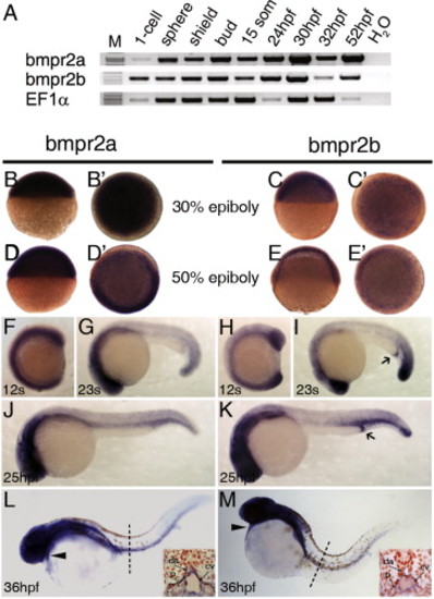
Expression of bmpr2a and bmpr2b in zebrafish development. (A) Analysis of bmpr2a and bmpr2b expression by RT-PCR. RNA for both receptors was detected from 1-cell stage embryos to 52 hpf. (B–E) In situ hybridization shows bmpr2a and bmpr2b are ubiquitously expressed at 30% and 50% epiboly. At 12 somite-stage, bmpr2a is broadly expressed throughout the embryo (F) whereas expression of bmpr2b is stronger in the most posterior and anterior regions (H). At 23 somites, both genes show a diffuse expression in the trunk and stronger staining in the head and tail regions (G, I). Bmpr2b is further expressed in the proctodeum at this stage (I, black arrow). (J) Bmpr2a is expressed in the head region, in the trunk vessels (dorsal aorta and cardinal vein) and in the posterior intermediate cell mass (ICM) at 25 hpf. (K) Bmpr2b is expressed in the head region and strongly in the posterior ICM and proctodeum (black arrow) but weakly in the trunk vessels. At 36 hpf, expression of bmpr2a (L) is restricted to the anterior region, including the heart (black arrowheads) and to the posterior blood vessels and pronephros (see inset). (M) Bmpr2b shows similar expression to bmpr2a, but it is not expressed in the dorsal aorta (see inset). (B–E) are lateral views and (B′–E′) are top views. (G, I–M) Lateral view, anterior to the left; (F,H) anterior to the top. Cv, cardinal vein, da, dorsal aorta, p, pronephros. |
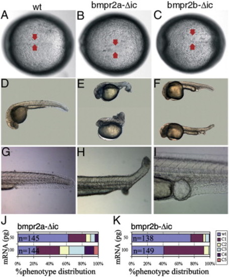
Injection of truncated bmpr2a and bmpr2b mRNA induce BMP loss-of-function phenotypes. Morphological phenotypes were classified according to Kishimoto and colleagues (Kishimoto et al., 1997). (A) Uninjected embryo at tailbud stage. Embryos were injected with 100 ng/μl of bmpr2a-Δic (B, E, H) or 100 ng/μl of bmpr2b-Δic (C, F, I). Injection of bmpr2a-Δic (B) or bmpr2b-Δic (C) induced an enlarged notochord at tailbud stage (red arrows). (D) Wild type embryo at 24 hpf. (E) Example of C2–C4 dorsalized phenotypes after injection of bmpr2a-Δic mRNA and (F) C2–C3 phenotypes after injection of bmpr2b-Δic mRNA. (G) Wild type tail at 24 hpf. (H) Partial tail duplication in bmpr2a-Δic-injected embryo. (I) Cloacal cysts appear frequently upon bmpr2a-Δic or bmpr2b-Δic injection. Anterior is to the left (A–I). Quantification of the number of embryos showing dorsalized phenotypes in each category (C1–C5) upon injection of 50 and 100 ng/μl bmpr2a-Δic (J) or bmpr2b-Δic mRNA (K). n - number of injected embryos at each concentration. |
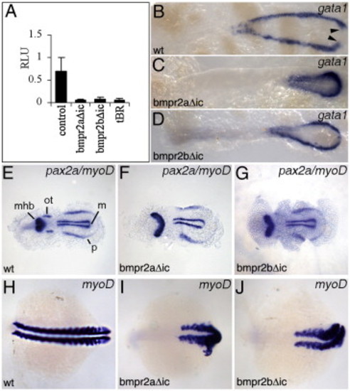
Overexpression of dominant negative bmpr2a and bmpr2b block BMP signalling in vivo. Unless otherwise stated, bmpr2a-Δic and bmpr2b-Δic mRNA were injected at 100 ng/μl. (A) Smad-dependent BMP signalling upon overexpression of full length or truncated bmpr2a and bmpr2b mRNA in embryos. Injection of 200 ng/μl of bmpr2a-Δic or bmpr2b-Δic mRNA efficiently blocked endogenous BRE-Luciferase activity in embryos at 70% epiboly. Injection of mRNA encoding tBR, a Xenopus dominant negative ALK3, was injected as a control. (B–D) Gata1 expression at 12 somites in wild type (B), bmpr2a-Δic (C) and bmpr2b-Δic (D) injected embryos. (E) At the 4-somite stage, pax2 is expressed in the midbrain–hindbrain boundary (mhb), otic placodes (ot) and pronephric (ventral) mesoderm (p), while myoD is expressed in the presomitic (axial) mesoderm (m) in wild type embryos. Embryos injected with bmpr2a-Δic (F) or bmpr2b-Δic (G) showed an expanded pax2 expression in the mhb domain concomitant with a strong reduction of pax2 expression in the otic placodes. (H) Expression of myoD in the somites of a wild type 14-somite stage embryo (lateral view, anterior to the right). Overexpression of bmpr2a-Δic (I) or bmpr2b-Δic (J) in 14-somite stage embryos. Note that the myoD expression is ventrally expanded, surrounding the most posterior end of the defective axis; the most anterior somites do not seem to be affected. RLU - Relative Luciferase Units. EXPRESSION / LABELING:
|

Morpholino (MO) knockdown of bmpr2a and bmpr2b affect normal cardiac jogging. (A) wild type embryo at 32 hpf. Knockdown phenotypes of bmpr2a morphants after injection with bmpr2aMO1 (B) and bmpr2aMO3 (C) and of bmpr2b morphants with bmpr2bMO1 (D) and bmpr2bMO3 (E). Gross morphology analysis shows a slight A–P axis shortening and cardiac oedema in bmpr2a and bmpr2b morphant embryos compared to wild type. The heart position in morphant and wild type sibling embryos at 32 hpf was scored as left jog (normal) (F), no jog (G) and right (reversed) jog (H). (I) Quantification of the heart jog defects observed in embryos injected with bmpr2aMO1, bmpr2aMO3, bmpr2bMO1 or bmpr2bMO3. Results are presented as the percentage of embryos which show a correct (left jog) or incorrect (no jog or right jog) heart jog after injection of the correspondent MO. The jog phenotypes were also scored in wild type, non-injected siblings and in embryos injected with the standard control MO. (J) Comparison of heart jog phenotypes induced by double bmpr2a/bmpr2bMO3 (0.25 mM + 0.05 mM) and single bmpr2aMO3 (0.25 mM) and bmpr2bMO3 (0.05 mM) injection. Single MO injections marked (*) are shown in panel I and J. Results are presented as the percentage of embryos which show a correct (left jog) or incorrect (no jog or right jog) heart jog after injection of the correspondent MO. The number (n) of injected embryos and the morpholino concentrations used are indicated in the graph. PHENOTYPE:
|

LR asymmetry of the heart and viscerae is perturbed in bmpr2a and bmpr2b morpholino-knockdown embryos. Analysis of cmlc2 expression at 32 hpf (A–G) and 48 hpf (H–N) in bmpr2aMO3 and bmpr2bMO3-injected embryos. (A) Wild type embryos show normal left-sided heart jog. Morpholino knockdown of bmpr2a (B, C) or bmpr2b (E–G) causes the heart to jog to the left, the right or stay at the midline. At 48 hpf, the linear heart tube has looped to the right (D-loop) and changed into an S-shaped tube (H). bmpr2a morphants show normal (I), no/reduced (J) or reversed looping (K). Similar results were found upon bmpr2b knockdown by bmpr2bMO3 (L–N). Comparison of foxa3 expression between wild type (O) and bmpr2a (P–R) or bmpr2b (S–U) morphants at 48 hpf showed that the direction of gut looping is also affected in these embryos. (A–G, O–U) Dorsal view, anterior to the top. (H–N) Frontal view, anterior to the top. |

Bmpr2a and bmpr2b regulate asymmetric expression of the left-specific genes lefty2, lefty1, spaw and pitx2 in the lateral plate mesoderm (LPM) and of bmp4 in the left heart field. (A) lefty2 is expressed at the 25-somite stage in the left heart field in wild type embryos. (B–E) Expression of lefty2 in bmpr2aMO3-, bmpr2bMO3- or bmpr2a/bmpr2bMO3-injected embryos (representative pictures, see Table 4). (F) lefty1 is expressed in the posterior notochord and left heart field at 22 somites. Bmpr2a-, bmpr2b- and double bmpr2a/bmpr2b morphants show normal (G), absent (H), bilateral (I) or right-sided (J) lefty1 expression in the heart field. (K) At 14–16 somites, spaw is expressed in the left LPM (arrowheads) while ntl expression marks the midline. (L–O) MO-induced knockdown of bmpr2a, bmpr2b or bmpr2a/bmpr2b (see Table 4) resulted in left (L), absent (M), bilateral (N) or rightward (O) spaw expression in the LPM. (P) In wild type embryos pitx2 is expressed in the left LPM. Bmpr2a, bmpr2b or bmpr2a/bmpr2b morphants display left-sided (Q), absent (R), bilateral (S) or reversed (T) pitx2 expression (arrowheads). (U) At 22 somites, bmp4 expression is higher in the left heart field. Bmpr2a, bmpr2b or bmpr2a/bmpr2b morphants display normal (V), symmetric (W) or reversed asymmetric bmp4 expression (X). (A–X) Dorsal view, anterior to the top. L, left, A, absent, B, bilateral, R, right. EXPRESSION / LABELING:
PHENOTYPE:
|
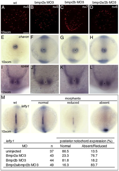
Analysis of cilia integrity in KV and of gene expression of charon, spaw and lefty1 at 10-somite stage in bmpr2 morphants. Anti α-acetylated tubulin staining of KV cilia in wild type (A), bmpr2a (B), bmpr2b (C) and bmpr2a/bmpr2b morphants (D) at 10 somites. (E) Perinodal expression of charon in wild type embryos. Expression was unchanged in bmpr2a (F), bmpr2b (G) or bmpr2a/bmpr2b morphants (H). Perinodal spaw expression at 12 somites showed no differences between wild type (I) and bmpr2a (J), bmpr2b (K) or bmpr2a/bmpr2b morphants (L). (M) Analysis of lefty1 expression at 10 somites in the posterior notochord of bmpr2 morphants. (A–D) Scalebar 50 μm. (E–H, M) Dorsal view, posterior to the top. (I–L) Dorsal view, anterior to the top. EXPRESSION / LABELING:
PHENOTYPE:
|

Bmpr2a and bmpr2b show different binding and Smad-dependent signalling transduction properties in vitro. (A) Co-immunoprecipitation of radioactive BMP2 ([125I]-BMP2) with overexpressed bmpr2a-myc, bmpr2b-Δic: :GFP and ALK3-HA in COS-7 cells. In the absence of ALK3, no binding of bmpr2a or bmpr2b to [125I]-BMP2 was detected. Bmpr2a but not bmpr2b co-immunoprecipitates with [125I]-BMP2 in the presence of ALK3-HA. (B) Direct western blot on cell extracts used for co-immunoprecipitation with antibodies against myc (bmpr2a), GFP (bmpr2b-Δic) and HA (ALK3) shows correct expression of the bmpr2a-myc, bmpr2b-Δic: :GFP and ALK3-HA constructs. (C) Transient transfection of bmpr2a, bmpr2b and bmpr2a + bmpr2b with or without BMP4 (50 ng/ml) in osteoblast progenitor cells. RLU, Relative Luciferase Units. These experiments have been repeated at least twice. |
Reprinted from Developmental Biology, 315(1), Monteiro, R., van Dinther, M., Bakkers, J., Wilkinson, R., Patient, R., Ten Dijke, P., and Mummery, C., Two novel type II receptors mediate BMP signalling and are required to establish left-right asymmetry in zebrafish, 55-71, Copyright (2008) with permission from Elsevier. Full text @ Dev. Biol.