
- Title
-
Genome-Wide Survey and Developmental Expression Mapping of Zebrafish SET Domain-Containing Genes
- Authors
- Sun, X.J., Xu, P.F., Zhou, T., Hu, M., Fu, C.T., Zhang, Y., Jin, Y., Chen, Y., Chen, S.J., Huang, Q.H., Liu, T.X., and Chen, Z.
- Source
- Full text @ PLoS One
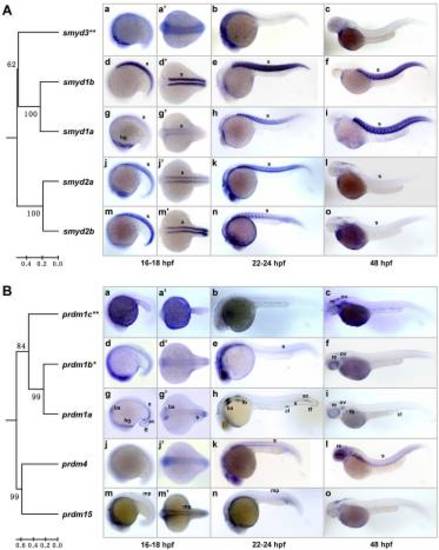
Somite/muscle-expressed SET domain genes and their evolutionary relationships. The phylogenetic relationships of the genes were indicated with the trees constructed based on the SET domains of the encoded proteins and rooted with zebrafish Smyd4 and Prdm14 proteins as outgroups, respectively. Lateral views (anterior to the left) of embryos at 16–18 hpf (a, d, g, j and m), 22–24 hpf (b, e, h, k and n) and 48 hpf (c, f, i, l and o) are presented. (a', d', g', j' and m') Dorsal views of the embryos in a, d, g, j and m. (A) Zebrafish smyd1a, smyd1b, smyd2a and smyd2b genes show somite/muscle-specific expression patterns and form a close paralog group with the smyd3 gene (double asterisks), which shows a ubiquitous expression pattern (a–c). Note the relatively low expressions of smyd1a at early stage (18 hpf; g) and smyd2a and smyd2b at late stage (48 hpf; l and o). (B) Expression patterns of the second paralog group. prdm1a is specifically expressed in anterior somites and adaxial cells at 18 hpf (g and g') and 24 hpf (h). Besides, it is also expressed in hatching gland (g), branchial arch, fin fold (g, g' and h), fin buds, cloaca (h and i) and retina (i). prdm1b (asterisk) is highly expressed in somites at 24 hpf (e) and in retina at 48 hpf (f). prdm1c (double asterisks) is ubiquitously expressed (a–c). prdm4 is highly expressed in somites and retina (k and l). prdm15 is expressed in muscle pioneer cells (m, m' and n). ac, adaxial cells; ba, branchial arch; cl, cloaca; fb, fin buds; ff, fin fold; hg, hatching gland; mp, muscle pioneer; re, retina; s, somite. EXPRESSION / LABELING:
|

Nervous system-expressed SET domain genes. (A) Expression patterns of closely related prdm3 and prdm16. (a–j) Lateral views (anterior to the left) of embryos at 12, 18, 24, 48 and 72 hpf. (b′ and g′) Ventral views of the embryos in b and g. (c′–e′ and h′–j′) Dorsal views of the embryos in c–e and h–j. Note the partially overlapping expression of prdm3 and prdm16. (B) Expression patterns of prdm13, prdm8a and prdm8b. (a–f) Lateral views (anterior to the left) of embryos at 24 and 48 hpf. (a′–f′) Dorsal views of the embryos in a–f. Note that the expression of prdm8a is mostly restricted in hindbrain and spinal chord (c, d and c′, d′), whereas that of prdm8b is restricted in olfactory placode, tegmentum, cerebellum and retina (f and f′). (C) Expression pattern of prdm12. (a and b) Lateral views (anterior to the left) of embryos at 18, 24 hpf. (a′ and b′) Dorsal views of the embryos in a and b. At 48 hpf, prdm12 is expressed in olfactory placode, tegmentum, cerebellum and hindbrain. (D) Expression pattern of prdm15. (a and b) Lateral views (anterior to the left) of embryos at 18, 22 hpf. (a′ and b′) Dorsal views of the embryos in a and b. Note that prdm15 is expressed in cranial ganglia neurons (a′ and b′) as well as in muscle pioneer cells and intermediate cell mass (a and b). ba, branchial arches; ce, cerebellum; cg, cranial ganglia; cnc, cranial neural crest; fb, fin buds; hb, hindbrain; icm, intermediate cell mass; mp, muscle pioneer; op, olfactory placode; pnd, pronephric duct; re, retina; sc, spinal chord; tel, telencephalon; tg, tegmentum; vd, ventral diencephalons. EXPRESSION / LABELING:
|
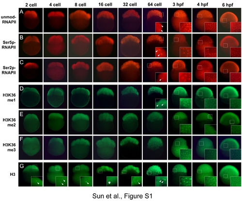
Immunofluorescent analyses of RNA polymerase II phosphorylation and histone H3K36 methylation in zebrafish embryos. Zebrafish embryos at different stages were subject to immunofluorescent staining to detect the unmodified pol II (A) and hyperphosphorylated pol II (B and C), H3K36 monomethylation (D), dimethylation (E) and trimethylation (F). Immunofluorescent staining of histone H3 (G) was used as a positive control. While the staining of histone H3 in nuclei is consistently detected (G), the staining of H3K36 methylation cannot be detected until 64-cell stage (D–F). The inset panels show the magnified views of detected staining in nuclei (arrow head). The unmodified, serine 2-phosphorylated and serine 5-phosphorylated pol II were probed with mouse monoclonal antibodies 8WG16, H5 and H14 (Covance Research Products), respectively. H3K36 mono-, di- and trimethylation were probed with rabbit polyclonal antibodies ab9084 (ABcam), 07-274 (Upstate) and ab9050 (Abcam), respectively. Histone H3 was probed with rabbit polyclonal antibody ab1791 (Abcam). |

Representative examples of ubiquitously expressed SET domain genes with relatively higher expression in certain tissues. Lateral views (anterior to the left) of embryos at 18 hpf (a, c, e and g) and 24 hpf (b, d, f and h) are presented. Note that whsc1 (a and b) and ezh2 (c and d) are highly expressed in the central nervous system, whereas ezh2 (c and d), setd2 (e and f) and mll5 (g and h) are highly expressed in intermediate cell mass of mesoderm. cns, central nervous system; icm, intermediate cell mass. EXPRESSION / LABELING:
|

Expression of SET domain genes before the onset of zygote gene transcription. WISH analyses of 58 zebrafish SET domain genes at 0.75, 2 and 4 hpf were representatively shown. EXPRESSION / LABELING:
|

Unillustrated author statements EXPRESSION / LABELING:
|