
- Title
-
AML1-ETO reprograms hematopoietic cell fate by downregulating scl expression
- Authors
- Yeh, J.R., Munson, K.M., Chao, Y.L., Peterson, Q.P., MacRae, C.A., and Peterson, R.T.
- Source
- Full text @ Development

Expression of AML1-ETO in zebrafish embryos causes an accumulation of hematopoietic cells. (A) Schematic diagram of the DNA fragment used to generate the Tg(hsp:AML1-ETO) zebrafish line. (B,C) Bright-light images of 1-dpf embryos that have been subjected to heat treatment. The accumulated hematopoietic cells in Tg(hsp:AML1-ETO) embryos are indicated with red arrows (C). The areas in the boxes are shown at a higher magnification in D and E. PHENOTYPE:
|

AML1-ETO transgenic zebrafish possess functional cardiovascular systems. (A) flk1 in situ hybridization of heat-treated wild-type and Tg(hsp:AML1-ETO) embryos harvested at designated stages as indicated. (B) Bright-light and fluorescent images of fli1-EGFP transgenic embryos in wild-type or Tg(hsp:AML1-ETO) background. These embryos have been subjected to the same heat treatment. The arrowhead indicates the site of the accumulated blood cells. (C) Fluorescent microangiography shows that the vasculature is continuous and the site of the accumulated blood cells is connected to the rest of the circulatory system. EXPRESSION / LABELING:
PHENOTYPE:
|

The accumulated hematopoietic cells in Tg(hsp:AML1-ETO) embryos are enriched for immature blast cells. (A-G) Cytology of hematopoietic cells from wild-type (A,D,E) and Tg(hsp:AML1-ETO) (B,C,F,G) embryos at 40 hpf. Low magnification (A,B); high magnification (C-G). Arrowheads, blast-like cells (C); black arrow, a bi-nucleated heterophil/neutrophil (D). |
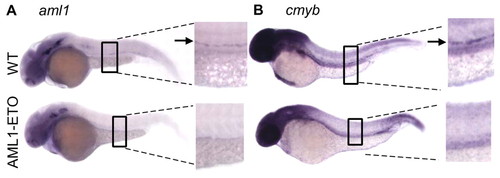
AML1-ETO disrupts definitive hematopoiesis. (A,B) In situ hybridization of (A) aml1 and (B) cmyb at 33 hpf indicates that definitive hematopoiesis is blocked in Tg(hsp:AML1-ETO) embryos. |

AML1-ETO reprograms hematopoietic cell fate, converting erythropoiesis to granulopoiesis. (A) In situ hybridization of gata1, fluorescent images of zpu.1-EGFP transgenic fish, and in situ hybridization of mpo and l-plastin. AML1-ETO expression results in gata1 downregulation. Subsequently, pu.1 expression is increased. Finally, the accumulated blood cells express the granulocytic cell marker mpo but not the monocytic cell marker l-plastin. All embryos were subjected to the heat treatment and then were collected at designated stages as indicated. (B) Proposed effects of AML1-ETO in hematopoietic progenitor cells. MPC, myeloerythoid progenitor cell; GMP, granulocyte/monocyte progenitor; MEP, megakaryocyte/erythroid progenitor. The red crosses indicate the steps suppressed by AML1-ETO. The parentheses indicate the markers for each cell type or the genes involved in the processes. These data suggest that AML1-ETO reprograms hematopoietic cell fate, resulting in an enrichment of myeloblasts that express mpo (boxed and shaded). EXPRESSION / LABELING:
|

AML1-ETO suppresses the monocytic cell fate. (A) In situ hybridization of l-plastin, mpo and pu.1. Tg(hsp:AML1-ETO) embryos were injected with gata1 morpholino (MO). Half of the injected embryos were heat-treated to induce AML1-ETO expression. AML1-ETO suppresses the expression of pu.1 and l-plastin but promotes the expression of mpo in gata1 morphants. (B) In situ hybridization of mpo. Injections of pu.1 MO decreased the expression of mpo in AML1-ETO-expressing embryos, inicating that Pu.1 is essential for the specification of granulocytes. EXPRESSION / LABELING:
|

AML1-ETO downregulates scl, leading to the early effects of AML1-ETO expression in primitive hematopoietic cells. (A) In situ hybridization of scl. AML1-ETO expression results in scl downregulation specifically in hematopoietic cells. The expression of scl in the head (asterisk), in the endothelial primordium (arrowhead), and in the posterior tail region (arrow) was not affected. (B) In situ hybridization of gata1, mpo and fluorescent images of zpu.1-EGFP transgenic zebrafish. Injections of scl mRNA restored gata1 expression and reversed pu.1 and mpo induction in Tg(hsp:AML1-ETO) embryos. EXPRESSION / LABELING:
|
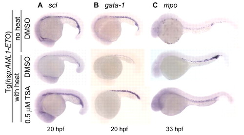
Trichostatin A suppresses the effects of AML1-ETO in zebrafish embryos. (A-C) DMSO or 0.5 µM TSA was added to Tg(hsp:AML1-ETO) embryos 2 hours before (A,B) or at the end of a 1-hour heat treatment (C) at 38°C at 18 hpf. Tg(hsp:AML1-ETO) embryos that had not been subjected to the heat treatment were used as a control. TSA treatment rescues scl and gata1 downregulation and blocks the accumulation of Mpo+ cells caused by AML1-ETO expression. EXPRESSION / LABELING:
|
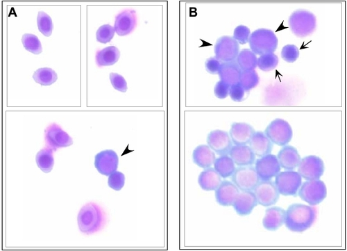
Transient expression of AML1-ETO results in accumulation of immature blast cells that fail to mature. (A,B) Cytology of hematopoietic cells from wild-type (A) and Tg(hsp:AML1-ETO) (B) embryos at 5 dpf. All of the embryos have been subjected to 1-hour 38°C incubation at 16 hpf. All of the wild-type fish exhibit abundant circulating blood cells at the time of harvest. Only some of the Tg(hsp:AML1-ETO) embryos possess circulating blood cells at the time of harvest. Pools of accumulating blood cells can still be seen in many of the Tg(hsp:AML1-ETO) embryos. (A) The wild-type sample was extracted from the circulating blood cells. It contains mostly mature erythrocytes, while immature blast cells (arrowhead) are scarcely seen. (B) The Tg(hsp:AML1-ETO) sample was extracted from the non-circulating blood cells. It contains a mixture of erythroid cells (arrows) and immature blast cells (arrowheads). |

The Mpo+ cells accumulated in Tg(hsp:AML1-ETO) embryos are unlikely to be immature erythroid progenitors. (A) Bright field and (B) fluorescent image of double in situ hybridization of Mpo (purple color, no fluorescence) and βe3 globin (red color, red fluorescence) at 31 hpf. (C) Combined image of both bright field and fluorescence image. The photos show the side view of the embryo close to the end of yolk extension (marked in blue in C). Two filled arrows point to the Mpo+ cells stained purple but have no red fluorecence, suggesting that these cells do not express βe3 globin. By contrast, the cell that expresses βe3 globin does not express Mpo (open arrow). EXPRESSION / LABELING:
|

AML1-ETO expression results in cebpa downregulation in hamatopoietic cells. In situ hybridization of cebpa indicates that in AML1-ETO transgenic embryos, hematopoietic expression of cebpa is blocked (red arrows), whereas intestinal expression of cebpa is unaffected (black arrows). Both embryos have been subjected to the same heat treatment. EXPRESSION / LABELING:
|
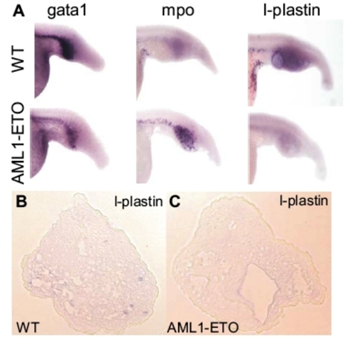
AML1-ETO suppresses the erythroid and monocytic cell fates, and promotes the granulocytic cell fate in chordin morphants. (A) In situ hybridization of gata1, mpo and l-plastin in wild-type and AML1-ETO transgenic embryos. All of the embryos have been injected with 50 mM chordin antisense morpholino oligonucleotides (Open Biosystems) and subjected to the same heat treatment. The expanded pool of blood cells in the chordin morphants expresses gata1 and l-plastin (A, upper panels), whereas induced expression of AML1-ETO downregulates both gata1 and l-plastin but upregulates mpo (A, lower panels). The embryos were at 22 hpf for gata1 staining and 31 hpf for mpo and l-plastin staining. (B,C) Cross sections of chordin morphants stained with l-plastin probes. While l-plastin+ cells are often seen in the wild-type embryos, these cells are not seen in the AML1-ETO transgenic embryos. EXPRESSION / LABELING:
|