
- Title
-
Six cadm/synCAM genes are expressed in the nervous system of developing zebrafish
- Authors
- Pietri, T., Easley-Neal, C., Wilson, C., and Washbourne, P.
- Source
- Full text @ Dev. Dyn.
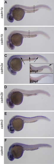
Expression of cadms at 24 hours postfertilization (hpf). In situ hybridization performed on 24 hpf whole-mount zebrafish embryos reveals that the six cadm genes are expressed throughout the central nervous system, in particular in the developing brain, the visual system and the spinal cord. A-C: Expression for cadm1a and 1b is in a rostrocaudal gradient (A,B), being undetectable caudal to the dashed lines. cadm2a expression decreases rostrally at 48 hpf and is lost by 72 hpf (insets in C). EXPRESSION / LABELING:
|
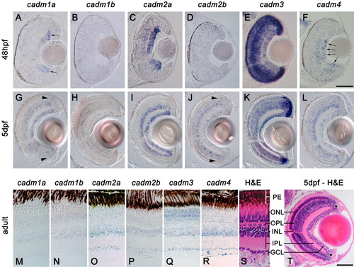
Expression of cadms in the visual system. In situ hybridization staining in sections of retina at 48 hpf (A-F), 5 days postfertilization (dpf; G-L), and adult zebrafish (M-R). S and T show hematoxylin and eosin staining (H&E) of sections of adult and 5 dpf, respectively; the different layers of the retina are indicated: PE, pigment epithelium; ONL, outer nuclear layer; OPL, outer plexiform layer; INL, inner nuclear layer; IPL, inner plexiform layer; GCL, ganglion cell layer. Asterisk in T denotes the location of the marginal zone. cadm1a is expressed in the dividing ganglion cells (arrows in A), while cadm4 is more strongly expressed in the mature cells of the GCL (arrows in F). Arrowheads in G and J reveal precursors at the margin of the outer nuclear layer (ONL). Scale bars = 50 μm in A-F, 50 μm in G-L,T. |
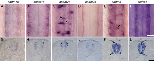
Expression of cadms in the developing spinal cord. A-L: The cadm expression revealed by in situ hybridization in whole-mount (dorsal view, A-F) and cross-sections (G-L) of 48 hpf embryos. Due to the strong rostrocaudal gradient of cadm1b expression, panels B and H show an anterior localized region of the spinal cord (somites 3-5), immediately behind the hindbrain, whereas all other images are from a region dorsal to the anus (somites 12-15). Staining for cadm2a and 3 is in dorsal cells at the midline of the spinal cord, suggestive of sensory Rohon-Beard neurons (arrowheads in C and E, arrow in K). Expression for cadm3 is also seen in the dorsal root ganglion (arrowhead in K). cadm4 is strongly expressed in the ventral domain of the spinal cord indicative of floor plate or motoneurons. Scale bars = 20 μm in A-F, 20 μm in G-L. EXPRESSION / LABELING:
|