
- Title
-
Expression of unconventional myosin genes during neuronal development in zebrafish
- Authors
- Sittaramane, V., and Chandrasekhar, A.
- Source
- Full text @ Gene Expr. Patterns

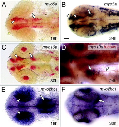
Myosins exhibiting restricted expression patterns in the hindbrain. All panels show dorsal views of the hindbrain with anterior to the left. Asterisks mark the otic vesicle in each panel. (A and B) myo10b is expressed at varying levels in rhombomeres 2–5 (A), and myo15a is expressed in r3 and r5 (B), with both expressed at the midbrain–hindbrain boundary (mhb). (C) myo1e1 is weakly but specifically expressed in a mesendodermal domain spanning the 2nd and 3rd somites (arrowhead). (D and G) At 18 hpf, myo1b2 is expressed in sensory ganglia (tg, ag) and a cluster of cells in r2 (arrowhead, D). By 36 hpf (G), there is prominent expression in cells (arrowheads) at the boundaries of r5 and r6. (E and H) At 18 hpf, myo5a is expressed in sensory ganglia, and in differentiating neurons (arrowheads, E) within every rhombomere. By 24 hpf (H), expressing cells form a continuous column along the neural tube (arrowheads). (F and I) At 18 hpf, myo10a is expressed in the sensory ganglia (tg), and in the branchiomotor neurons in r2 and r5 (arrowheads, F). By 30 hpf (I), expression persists in the sensory ganglia and migrating motor neurons (arrowhead), and has expanded to large clusters of cells in the anterior hindbrain. tg, trigeminal ganglion; ag/pg, anterior and posterior lateral line ganglion. Scale bar, 75 μm. EXPRESSION / LABELING:
|
Myosin expression in the forebrain and midbrain. All panels show dorsal views of the head with anterior to the left. (A and B) At 18 hpf, myo5a is expressed in nascent neurons (arrowhead, A) in the forebrain, and in the nucMLF neurons (arrow) in the midbrain. By 24 hpf (B), forebrain expression has refined into distinguishable clusters representing the neurons of the anterior and post-optic commissures (arrowheads, B), and is maintained in the nucMLF neurons (arrow). (C) At 30 hpf, myo10a is expressed in the neurons of the nucMLF (arrow), and of the anterior and post-optic commissures (arrowheads), and in the lens (see Fig. 6). (D) Double-labeling shows that tubulin antibody-labeled axons (arrowhead) of the MLF arise from the cluster of myo10a-expressing neurons (arrow), corresponding to the nucMLF. (E and F) At 18 hpf, myo2hc1 is expressed in the medial layer of the optic cup (arrowheads), and at the midbrain–hindbrain boundary (arrow). By 32 hpf (F), expression is restricted to the retinal ganglion layer (see Fig. 6), and to the caudal edge of the developing optic tectum (arrow). Scale bar, 25 μm (D), 50 μm (A–F). EXPRESSION / LABELING:
|

Myosin expression in the developing ear. Upper panels show dorsal views, with anterior at top. Lower panels show cross-sections (dorsal up) at the levels indicated in the upper panels. (A and C) myo10b is expressed in epithelial cells (arrowhead) lining the anterior third (A), and the ventral half (C) of the otic vesicle. (B and D) myo2hc1 is expressed in the epithelial cells of the otic vesicle (arrowheads), adjacent to the hindbrain. Asterisks (C and D) indicate the notochord. Scale bar, 30 μm. EXPRESSION / LABELING:
|

Myosin expression in the eye. (A and B) myo1b2 is expressed in the differentiating lens fibers (arrowhead, A) located in the medial half of the lens (B, dorsal view). (C) myo10a is expressed in a subset of lens cells at 24 hpf, and at 30 hpf (see Fig. 4C). (D) myo2hc1 is expressed in retinal ganglion cells (arrowhead) adjacent to the lens. Asterisks (A and C) indicate the choroid fissure. Scale bar (A, B, and D), 40 μm. Scale bar (C), 30 μm. |

Myosin expression in the trunk and spinal cord. (A–C, E, G, and I) Side views with anterior to left. (D, F, H, and J) Cross-sections at levels indicated in (C, E, G, and I), respectively. (A) myo2hc2 is strongly expressed in the differentiating muscles of the somites (white arrowheads), but only weakly in the newly formed somites (black arrowhead). (B) myo18a is expressed in the mesenchyme of newly formed and differentiating somites (black arrowhead), and strongly in the cells at the boundaries of older somites (white arrowheads). (C and D) myo1b2 is expressed broadly in the trunk, excluding the presomitic mesoderm (C). Expressing cells are located superficially within the somitic mesoderm (arrowhead, D), and in the notochord (asterisk). (E and F) At 18 hpf, myo5a expression is restricted to the spinal cord at anterior trunk levels (E), and represents differentiating neurons (arrowhead, F). (G and H) At 30 hpf, myo5a is expressed by scattered cells located in the dorsal half of the spinal cord (G). A cross-section in the anterior trunk (H) shows that expressing cells include neurons (arrowhead) and migrating neural crest cells (arrows). (I and J) myo10a is expressed in a continuous column of cells in the dorsal half of the spinal cord (I), including neurons (arrowheads, J). Asterisks in (D, F, H, and J) indicate the notochord. Scale bar (I), 50 μm (A–C, E, G, and I). Scale bar (J), 50 μm (D, F, H, and J). EXPRESSION / LABELING:
|

Myosin2 expression in head muscles. (A) In a dorsal view, some eye (so, sr, lr) and jaw (lap) muscles can be identified. (B) In a side view, several eye (so, sr, mr, io, ir, am) and jaw (ah, ao, do, lap) muscles can be distinguished. (C and D) In a ventral view, several eye (am, io, ir, lr, mr), jaw (ima, imp, ih, hh) and gill (tv, sh) muscles can be identified. Scale bar, 100 μm (A–C), 50 μm (D). EXPRESSION / LABELING:
|
Reprinted from Gene expression patterns : GEP, 8(3), Sittaramane, V., and Chandrasekhar, A., Expression of unconventional myosin genes during neuronal development in zebrafish, 161-170, Copyright (2008) with permission from Elsevier. Full text @ Gene Expr. Patterns