
- Title
-
Cadherin-mediated adhesion regulates posterior body formation
- Authors
- Harrington, M.J., Hong, E., Fasanmi, O., and Brewster, R.
- Source
- Full text @ BMC Dev. Biol.

Loss of N-cadherin function causes axis shortening. (A-E) Lateral views of live 30 hpf zebrafish embryos imaged with Nomarski optics. Anterior is to the left, dorsal is up. Insets show images of somites at the level of the yolk sac extension. Black arrowheads point to brain defects observed in N-cad mutants. Black arrow points to the characteristic club-shaped, shortened tail in N-cadm117 homozygous mutants. WT (A), N-cadp79emcf homozygous mutant (B), N-cadr2.10 homozygous mutant (C), N-cadm117 homozygous mutant (D), N-cadm117/p79emcf transheterozygote mutant (E) embryos. PHENOTYPE:
|

Length of tail is not altered in N-cadm117/+ heterozygotes. Measurements of tail length of WT and offspring from a cross between WT and N-cadm117/+ heterozygous embryos at 30 hpf. Tail lengths of N-cadm117/+ heterozygotes were comparable to those in WT embryos. PHENOTYPE:
|
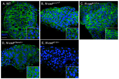
Expression and localization of N-cad protein in N-cad mutants. Cross-sections through the head region of 7 som stage WT (A), N-cadm117 homozygous mutant (B), N-cadm117 heterozygous mutant (C), N-cadp79emcf homozygous mutant (D) and N-cadr2.10 homozygous mutant (E) embryos, labeled with α-N-cad (green) and DAPI (blue). Insets show high magnification of cross-sections through the tailbud region of these same embryos. Scale bar, 20 μm. EXPRESSION / LABELING:
PHENOTYPE:
|
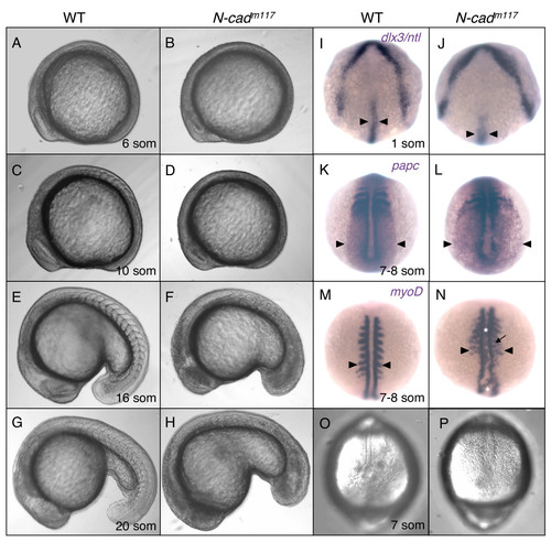
Mesodermal morphogenesis defects in N-cadm117 mutants. (A-H) Lateral views of WT (A,C,E,G) and N-cadm117 mutant (B,D,F,H) embryos imaged with Nomarski optics at 6 som (A,B), 10 som (C,D), 16 som (E,F) and 20 som (G,H). Anterior is to the left, dorsal is up. (I-N) Dorsal view of WT and N-cadm117 mutant embryos at 1 som (I,J) and 7–8 som (K-N) processed by in situ hybridization. (I,J) Dorsal anterior view of dlx3 and ntl expression. Black arrowheads indicate width of NC. (K,L) Dorsal posterior view of papc expression. Black arrowhead point to the lateral edge of the paraxial me3Edlx3 and ntl expression. Black arrowheads indicate width of NC. (K,L) Dorsal posterisoderm. (M,N) Dorsal view of myoD expression. Black arrowheads indicate length of somites. White asterisks indicate ectopic labeling in the axial mesoderm. Black arrow points to ectopic intersomitic myoD labeling. (O-P) Dorsal posterior view of WT (O) and N-cadm117 homozygous mutant (P) at 7 som, imaged with Nomarski optics. Abbreviations: som, somite. EXPRESSION / LABELING:
PHENOTYPE:
|

Expression of N-cadherin during gastrulation and somitogenesis. N-cadherin mRNA expression in WT, 60% epiboly (A-A″), 95% epiboly (B-B″), 3 som (C-C″), 6 som (D-D″), and 18 som (E-E″) embryos. Animal views (A,B), dorsal views (A′,B′), and sagital views (A″,B″). Asterisk indicates lack of N-cad expression in the ventral epidermis. (C-E″) Cross-sections through the head (C,D,E), trunk (C′,D′,E′), and tail (C″,D″,E″) regions. Insets indicate the angle at which the embryo was sectioned. Abbreviations: V, ventral; L, lateral; D, dorsal; A, animal; NT, neural tube; NC, notochord; som, somite; TB, tailbud; OV, otic vessicle; PSC, postmigratory slow cells. EXPRESSION / LABELING:
|

Impaired movement of anterior tailbud cells in N-cadm117 mutants. Posterior (A-C″) and anterior (D-G&prime) cells in the tailbud were uncaged at 4 som (A-C,D-G) and imaged at 18 som (B′-C″,E′-G′). (A,D) Schematic diagrams of a dorsal view of the tailbud at 4 som, illustrating where the uncaging was done (green dot). (B,C,E-G) Dorsal views of the tailbud at 4 som, showing where the uncaging was done (green label), in WT (B,E) and N-cadm117 mutants (C,F,G). Lateral (B′,E′) and dorsal (B″,E″) views of 18som WT embryos indicating the position of uncaged cells (white arrowheads). Lateral (C′,F′,G′) and dorsal (C″) views of N-cadm117 mutants indicating position of labeled cells (white arrowheads). Abbreviations and symbols: som, somite; kv, Kupffer′s vesicle; asterisk indicates the position of the vacuole in the tailbud, black arrowheads show KV. PHENOTYPE:
|
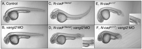
Genetic interaction between N-cadherin and vang-like 2. Lateral views of 30 hpf live embryos imaged using Nomarski optics. Anterior is to the left, dorsal is up. Control uninjected (WT) (A), WT injected with vangl2 MO (0.8 ng) (B), N-cadp79emcf homozygous mutant (C), N-cadp79emcf mutant injected with vangl2 MO (0.8 ng) (N-cadp79emcf; vangl2 MO) (D), N-cadm117 homozygous mutant (E), N-cadm117 mutant injected with vangl2 MO (0.8 ng) (N-cadm117; vangl2 MO) (F) embryos. Insets show images of somites at the level of the yolk sac extension. PHENOTYPE:
|
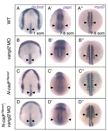
N-cadp79emcf; vangl2 MO embryos do not exhibit early gastrulation defects. Dorsal views of WT (A-A″), vangl2 MO-injected (B-B″), N-cadp79emcf homozygous mutant (C-C″), and N-cadp79emcf; vangl2 MO (D-D″) embryos labeled with dlx3 and ntl at 1 som (A-D), papc at 7 som (A′-D′), and myoD at 7 som (A″-D″). Black arrowheads indicate the width of the NC (A-D), the lateral edge of the paraxial mesoderm (A′-D′), and the length of the somites (A″-D″). Abbreviations: som, somite; NC, notochord. EXPRESSION / LABELING:
PHENOTYPE:
|
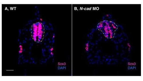
Loss of N-cadherin causes posterior neural tube defects. Cross sections through the posterior domain of the yolk sac extension of 30 hpf WT (A) and N-cad morpholino-injected (0.8 ng) (B) embryos labeled with α-Sox3C (pink) and DAPI (blue). Dotted line delineates the shape of the NT. Scale bar, 20 μm. |

vangl2 and kny do not regulate N-cad expression or localization. Cross-sections through the tail region of 18 som embryos labeled with α-N-cad (green) and DAPI (blue). N-cad is localized at the plasma membrane in WT (A),tri (B) and kny mutant (C) embryos. Insets show a higher magnification of N-cad labeling in the mesoderm. Dotted white circles show the location of the notochord. Scale bar, 20 μm. EXPRESSION / LABELING:
|
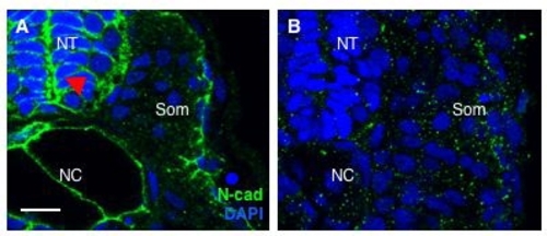
N-cad MO prevents translation of N-cad protein. Cross-sections through the tail region of 30 hpf embryos labeled with α-N-cad (green) and DAPI (blue). In WT, uninjected embryos (A) N-cad protein is expressed throughout the neural tube, where it is enriched at the apical surface (red arrowhead). In addition, N-cad is observed in the notochord and postmigratory slow cells (PSCs). Labeling is absent in the N-cad morpholino-injected (0.8 ng) (B) embryos. Abbreviations: NT, neural tube; som, somite; NC, notochord. Scale bar, 10 μm. EXPRESSION / LABELING:
|

Unillustrated author statements |