
- Title
-
The INT6 Cancer Gene and MEK Signaling Pathways Converge during Zebrafish Development
- Authors
- Grzmil, M., Whiting, D., Maule, J., Anastasaki, C., Amatruda, J.F., Kelsh, R.N., Norbury, C.J., and Patton, E.E.
- Source
- Full text @ PLoS One
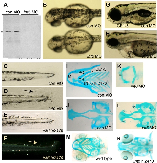
Int6 is essential for zebrafish embryonic development. (A) Western blot analysis of Int6 (*) in zebrafish embryos injected with a control (con) MO or int6 MO. (B) int6 morphant melanocytes are less darkly pigmented. (C–F) Int6 is required for pigment cell placement in the tail, as int6 morphants and int6hi2470 mutants have misplaced pigment cells in the tail fin (D, arrow). Ambient light illuminates the iridophore ‘star-light’ pattern seen in the int6hi2470 embryos (F, arrow). (G–H) By 5 dpf, embryos injected with a con MO have clearly visible certobranical arches, while int6 morphants do not have visible certobranical arches, in addition to other abnormalities, including unconsumed yolk sac, heart and eye development. (I–N). Alcian blue staining of 5 dpf embryos shows loss of ceratobrancial arches 1 through 5. M, Meckel's; PQ, palatoquadrate; CH, ceratohyal; CB, ceratobrancial. PHENOTYPE:
|
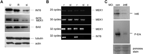
INT6 is required for MEK protein levels and Erk-signaling. A. Osteosarcoma U2-OS cells untransfected (U), or transfected with siRNA targeted against the INT6 mRNA (si) or the reverse sequence (R), show reduced levels of INT6 and MEK1 protein specifically after transfection with INT6-siRNA, but no reduction in BAX, tubulin or actin protein levels. (B) Semi-quantitative-PCR shows MEK1 mRNA is unaffected in reverse sequence and INT6-siRNA treated cells, coupled with the expected reduced levels of the INT6 message in the INT6-siRNA transfected cells. c, PCR control without DNA. (C) Phospho-Erk levels are reduced in int6 morphants, while ponceau stain detects equal loading of protein on the gel. |

Int6 and phospho-Erk expression in the developing zebrafish embryo, and pharmacological inhibition of Mek alters ceratobrancial (CB) arches.(A–F). Immuno-histochemistry of Int6 and phospho-Erk in the developing craniofacial tissues, counter stained with hematoxylin, and phospho-histone H3 to show cycling cells. (G, H) Ventral whole mount views of Alcian blue stained pharyngeal cartilages show loss of ceratobrancial arches 1–5 and a reduction of Meckel's (M), palatoquadrate (PQ) and ceratohyal (CH) cartilages in 4 dpf embryos treated with 0.5 uM CI-1040. (I, J) Sections of 4 dpf embryos hematoxylin and eosin stained after 0.5 uM CI-1040 treatment reveals loss of CB arches 1–5 (brackets). E, Ethmoid plate; PC, Parachordal cartilage. EXPRESSION / LABELING:
|

Int6 and Mek signaling interact in vivo. (A–C). Only embryos treated with the 1.0 μM CI-1040, and not 0.25 μM, show a loss of posterior structures, in contrast to (D) int6 morphants (int6 MO 0.25 ng). (E, F) In combination with 0.25 μM of CI-1040, 0.25 μg of int6 MO causes a dramatic alteration of the anterior-posterior axis. |
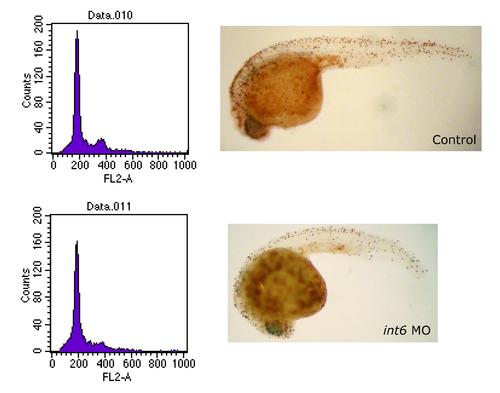
Cell cycle analysis of int6 morphants. Whole-mount immunohistochemistry with the late G2/M phase marker, phospho-histone H3 shows only slightly reduced numbers of cells in late G2/M phase in the int6 morphant compared to the control. Similarly, DNA content as measured by flow cytometry reveals only a slight reduction of cells in G2/M phase in the int6 morphant. Thus, we find that loss of Int6 in normal vertebrate cells (as well as in additional human cancer cell lines, M.G. & C.J.N. unpublished data) does not appear result in an accumulation of cells in G2/M progression. |

Lateral views of in situ hybridization of neural crest markers in control and int6 morphants, revealing no change in cell number or migration as indicated by the apparently normal expression of dlx2 (stages 6–36 hpf, examined at two hour intervals), nor of early markers of NCC and melanocytes, such as sox10, crestin, snail and mitfa (24 hpf) in int6 morphants. These observations were extended by examination of a transgenic sox10-GFP line (1) revealing unaltered GFP-expressing NC-derived cells in int6 morphants within the first 48 hpf, but a loss of GFP expressing differentiated pharyngeal arches 3–7 by 3 dpf (data not shown). |
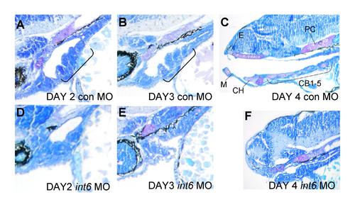
Development of the pharyngeal arches in the developing control (A–C) and (D–F) int6 morphant animals. Note the loss of pharyngeal arches (A, bracket) in the int6 morphants (bracket). Sections were stained with methylene blue. Anterior to the left. PHENOTYPE:
|
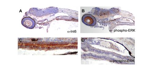
Immunohistochemistry of Int6 and phospho-Erk staining in 4 dpf embryos. (A, B) While Int6 and phospho-Erk signaling overlap in the craniofacial region, they also have distinct patterns, for example in the eye and (C, D) gut. We note that while Int6 and phospho-Erk have overlapping domains of expression in the craniofacial region, Int6 staining in the craniofacial region was stronger than phospho-Erk, and phospho-Erk staining was limited to specific tissues within the craniofacial region. M, Meckel′s; E: Ethmoid plate; CH, ceratohyal; CB, ceratobrancial. Sagittal section, anterior to the left. |