
- Title
-
A global role for zebrafish klf4 in embryonic erythropoiesis
- Authors
- Gardiner, M.R., Gongora, M.M., Grimmond, S.M., and Perkins, A.C.
- Source
- Full text @ Mech. Dev.

Anemia and impaired erythroid cell differentiation in klf4 morphants. Anemia and pericardial oedema in klf4-MO injected embryos (B) compared with Std-MO injected controls (A). May–Grunwald–Giemsa stained blood smears show a mixture of immature (top row) and maturing (bottom row) primitive erythroid cells in Std-MO injected controls (C) but only immature erythroid cells in klf4-MO injected embryos (D). PHENOTYPE:
|
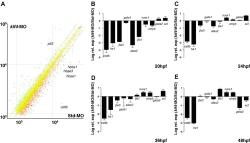
A global role for klf4 in embryonic erythropoiesis and hatching gland gene expression. (A) Scatter plot of mean relative expression (from four biological replicates) in klf4-MO injected embryos (y-axis) versus Std-MO injected embryos (x-axis). Lines representing 1.4-fold up and down regulation are shown. A few selected differentially expressed genes are indicated. (B–E) Quantitative real time-PCR validation of gene expression for catL, he1, βe3-globin, gata1, alas2, βa1-globin, runx1, c-myb, gata2, and scl at 20 hpf (B), 24 hpf (C), 36 hpf (D) and 48 hpf (E). Values are determined from the delta CT value and expressed relative to expression of γ-actin. Values are means of four experiments and are expressed as log relative expression of klf4-MO divided by Std-MO. EXPRESSION / LABELING:
|

Reduced embryonic βe3 globin and alas2 gene expression but not gata1, gata2, or scl gene expression in klf4 morphants. Whole mount in situ hybridization at 24 hpf for expression of the indicated genes. (A–D, I–L) klf4-MO injected embryos and E–H, M–N are Std-MO injected controls. In A–H dual in situ hybridization has been performed with catL to confirm loss of the hatching gland, and a specific probe as indicated. Arrows indicate the ICM and arrowheads the presence or absence of the hatching gland. |

Organisation of CACCC, TATA, and CCAAT box motifs in zebrafish and human globin genes and CACCC box binding by Klf4. (A) Schematic of the organisation of the zebrafish embryonic (e) and adult (a) α and β globin genes on chromosome 3. (B) Organisation of the zebrafish βe1, αe1 and βa1 globin gene promoter elements derived from BX004811. The human &epsilon-, &gamma-, and β-globin gene promoter elements were derived from U01317. The duplicated human γ-globin genes, Gγ and Aγ, have identical promoter sequences. Numbers indicate spacing in base pairs between the TAAA, CACCC and CCAAT box elements. Arrows indicate canonical KLF binding sites. Bases in bold indicate bases which differ from the 9 bp consensus, CCN–CNC–CCN. (C) Electromobility gel shift assay showing both KLF1 and Klf4 bind to the human β globin CACCC site (lanes 3 and 5). In addition, Klf4 binds to the embryonic βe1 globin CACCC site (lane 8) and a non-canonical CACCC site in the βa2 globin promoter (lane 14) indicated by a gel supershift in the presence of a V5 antibody (lanes 9 and 15). Klf4 does not bind the αe1 globin CACCC site (lanes 11 and 12). The motilities of SP1, SP4, and SP3 DNA complexes from COS7 cells were assigned based on previous work using specific antibodies to these. |

Unillustrated author statements |
Reprinted from Mechanisms of Development, 124(9-10), Gardiner, M.R., Gongora, M.M., Grimmond, S.M., and Perkins, A.C., A global role for zebrafish klf4 in embryonic erythropoiesis, 762-774, Copyright (2007) with permission from Elsevier. Full text @ Mech. Dev.