
- Title
-
The role of maternal Activin-like signals in zebrafish embryos
- Authors
- Hagos, E.G., Fan, X., and Dougan, S.T.
- Source
- Full text @ Dev. Biol.

Embryos lacking maternal sqt function have variable defects. Lateral (A, C–E, G, I, K) or ventral (B, D, F, H, J, L) images of live embryos at 30 hpf. The body axis forms normally in wild type (A), Zsqt (C) and Msqt (E) embryos, and none of these embryos are cyclopic (B, D, F), although Zsqt mutants from some crosses display mild cyclopia. Embryos lacking both maternal and zygotic sqt can be classified into three groups depending on their phenotype. The frequency of the phenotypes depends on the genetic background of the mother, as described in the text. The MZsqt embryos depicted in panels G–L are siblings. Class I MZsqt mutants (G, H) are indistinguishable from wild type, while Class II mutants (I, J) display mild cyclopia (J), typical of the previously reported phenotype of Zsqt mutants (Dougan et al., 2003). Class III mutants have reduced dorsal and anterior structures, characterized by reduced (L, arrow) or absent eyes. Anterior is to the left when apparent. PHENOTYPE:
|

Dorsal and anterior neural tissue is reduced or missing in Class III MZsqt embryos. emx1 (A–E) or pax6a (F–J) expression in wild type (A, F), Class I MZsqt (B, G), Class II MZsqt (C, H), Class III MZsqt (D, I), or MZoep (E, J) embryos at 24 hpf. emx1 expression in the posterior telencephalon is indistinguishable from wild type in Class I and Class II MZsqt (A–C), but is expanded in Class III MZsqt (D) and in MZoep (E). pax6a is expressed in the retinas of wild type and Class I MZsqt embryos (F, G) and in the retina of the cyclopic eyes in Class II MZsqt (H) and MZoep (J). pax6a is not expressed in Class III MZsqt embryos (I). (A–E) Lateral view with anterior to the left. (F–J) Dorsal view with anterior to the left. Arrowheads indicate retinal pax6a expression (F–H, J) or the absence of retinal pax6a expression (I). |

Maternal Activin-like signals are not required during the cleavage stages. Images of live embryos at 24 hpf. Embryos were treated with DMSO (A, B) or with SB-505124 at 0.75 hpf (C, D, G–L) or at 3 hpf (E, F). In panels G–L, the drug was applied at 0.75 hpf and washed out at the indicated time. Embryos treated with SB-505124 at 0.75 hpf and 3 hpf (C, E) lack all derivatives of the mesoderm and endoderm in the head and trunk, and are severely cyclopic (D, F). (G, H) When embryos are exposed to the drug only between 0.75 hpf and 3 hpf, they are indistinguishable from wild type (A, B). (I, J) When embryos are exposed to SB-505124 between 0.75 hpf and 4.3 hpf, they lack head mesoderm and endoderm, as indicated by severe cyclopia (J), but contain trunk somites and notochord (I). Finally, embryos exposed to SB-505124 between 0.75 hpf and 6 hpf lack notochord, but contain trunk somites (K), and are severely cyclopic (L). Lateral views, anterior is to the left (A, C, E, G, I, K) or ventral views (B, D, F, H, J, L). |
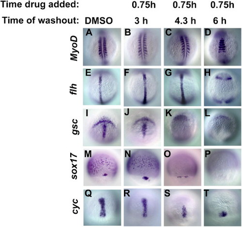
Progressive loss of cell fates with shorter exposures to Activin-like signals. Embryos were treated with DMSO (A, E, I, M, Q) or with SB-505124 between 0.75 hpf and 3 hpf (B, F, J, N, R), 4.3 hpf (C, G, K, O, S) or 6 hpf (D, H, L, P, T). Sibling embryos were divided into five groups. One group was fixed at 14 hpf and stained for MyoD expression (A–D). Embryos fixed at 10 hpf were stained for either flh (E–H), or gsc (I–L). Embryos fixed at 8 hpf were stained for either sox17 (M–P) or cyc (Q–T). When the drug is washed out at 3 hpf (B, F, J, N, R), expression of all markers is indistinguishable from wild type (A, E, I, M, Q). When the drug is washed out at 4.3 hpf, MyoD and flh expression is normal (C, G), but gsc is absent from the prechordal plate (K) and sox17 is only expressed in the dorsal forerunner cells (O). The axial domain of cyc expression is reduced (S). When the drug is washed out at 6 hpf, MyoD is expressed in trunk somites (D), and flh is expressed in four ectodermal domains, but not in the axial mesoderm (H). gsc is absent from the prechordal plate (L) and sox17 expression is abolished (P). cyc expression in the axial mesoderm is severely reduced, but is still present (T). In panels A–L, images are dorsal views, with anterior to the top of the page. In panels M–I, images are dorsal views with the animal pole at the top. EXPRESSION / LABELING:
|
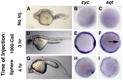
oep function is not required before 3 hpf. Images of uninjected MZoep mutants (A–C), or those injected with 100 pg sOep mRNA into the YSL at 3 h (D–F) or 4 h (G–I). MZoep embryos lack all derivates of mesoderm and endoderm in the head and trunk at 24 h (A), and lack expression of cyc (B) and sqt (C) at 5 hpf. Injection of sOep mRNA at 3 hpf restores mesoderm and endoderm (D), as well as cyc (E) and sqt (F) expression. Embryos injected at 4 h are severely cyclopic, but contain trunk somites and notochord (G). Expressions of both cyc (H) and sqt (I) are severely reduced. Animal pole views, with dorsal to the right. |

Treatment with SB-505124 after MBT enhances the MZoep mutant phenotype. MZoep mutants were treated with DMSO (A, C), SB-431542 at the 16- to 32-cell stage (B), or SB-505124 at 3 hpf (D). The majority of MZoep mutants treated with DMSO develop long tails containing somites (A), but a minority has underdeveloped tails with no apparent somites (C). After treatment with SB-431542 during the cleavage stages, all MZoep mutants have shortened tails with no apparent somites (B). Similarly, MZoep mutants have short tails after treatment with SB-505124 at 3 hpf (D). Images of live embryos at 24 h, anterior to the left. |
Reprinted from Developmental Biology, 309(2), Hagos, E.G., Fan, X., and Dougan, S.T., The role of maternal Activin-like signals in zebrafish embryos, 245-258, Copyright (2007) with permission from Elsevier. Full text @ Dev. Biol.