
- Title
-
Control of morphogenetic cell movements in the early zebrafish myotome
- Authors
- Daggett, D.F., Domingo, C.R., Currie, P.D., and Amacher, S.L.
- Source
- Full text @ Dev. Biol.

Adaxial cells undergo a series of stereotypical behaviors as the zebrafish myotome matures. Confocal images of adaxial cells at the depth of the notochord within 8–10 somite stage embryos labeled with phalloidin (green) and DAPI (red). Dorsal view, anterior to the left in all panels. (A) Schematic diagram of an 8-somite stage embryo highlighting the regions where subsequent panels B–F are centered along the axis. (B) Adaxial cells in the posterior presomitic mesoderm region appear cuboidal (arrowheads) and become more columnar with actin-rich apices in anterior presomitic mesoderm (arrows). (C) As somite boundaries begin to form, adaxial cells begin to lose the columnar shape, displaying more irregular shapes (arrowheads) before becoming more rounded and flattened, and interleafing with one another within the dorsoventral aspect of the forming somite (arrow). (D) As the adaxial cells begin to intercalate, they transiently display actin-rich apical constrictions, with the apices of 2–4 adjacent adaxial cells apparently interacting with one another and with the more lateral cells in the central somite (arrow). (E) Following the release of the apical interactions, adaxial cells continue to broaden, as the number of adaxial cells in a given dorsal ventral section of the somite is ultimately reduced to one. Arrow shows two broad adaxial cells with interacting membranes midway between somite boundaries. Asterisks point out that as somites mature, the nuclei of broadening adaxial cells become superimposed as the adaxial cells ultimately form one dorsoventral stack of cells in the medial somite. (F) Fully intercalated adaxial cells attached to somite boundaries in the anterior somites of an 8–10 somite embryo. (G) Arrows highlight another example of the transition from early interleafing stage adaxial cells with actin-rich apices, to triangular cells displaying apical constrictions shown across three adjacent somites. (H) Broad, intercalated adaxial cells with thin, lamellar lateral membranes appear to maintain membrane interactions with more lateral somitic cells (arrowheads), and display actin-rich, bipolar somite border attachments (arrows). (I) Schematic summary of the stereotypical behaviors displayed by adaxial cells prior to their migration. |

Adaxial cell behaviors occur with a dorsal to ventral progression within the developing myotome. Anterior to left in all panels. (A) In a more dorsal plane of adaxial cells adjacent to the notochord, the more anterior cells are broadening (*) while more posterior cells are undergoing interleafing behaviors (arrows). (A2) In a slightly more ventral plane, the anterior adaxial cells are beginning to lose epithelial shape and to accumulate cortical actin, characteristic of the early interleafing stage (*), while the more posterior adaxial cells still predominantly display a pseudo-epithelial appearance (arrows). (B) A lateral image of 3 adjacent somites, focused just lateral to the notochord surface, shows that, in each somite, the more dorsal adaxial cells in the somite have undergone further elongation and intercalation than those more ventral. Arrowheads highlight somite boundaries. (C) Schematic representation of panel B, illustrating both that the majority of more dorsal cells within the myotome are further advanced in their behaviors than the more ventral, and further, that within a given dorsoventral plane, the adaxial cells in more anterior somites are more advanced in their behaviors than those in more posterior somites as is apparent in Fig. 1. A, anterior; P, posterior; D, dorsal; V, ventral. |
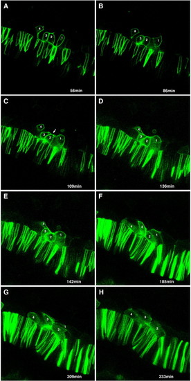
Time-lapse analysis of adaxial cell behaviors. Selected frames from Supplementary Movie. Four adaxial cells expressing a membrane-targeted GFP mRNA, that will eventually become incorporated into two adjacent somites, were followed for 4.5 h. Elapsed time, in minutes, of selected images shown. (A) Adaxial cells (1–4) are initially predominantly pseudo-epithelial, with their basal membranes against the notochord. Columnar shape is lost as adaxial cells become more irregularly shaped and rounded as they begin to interleaf (B), before beginning to make transient apical constrictions (arrow, C). (D–F) Adaxial cell membranes then begin to spread out as cells begin to broaden, with cell 1 showing a broad, lamellar expansion and cell 2 dramatically rounding (E) prior to further elongating. (F, G) Cells maintain broad character and laterally directed lamellar protrusions as they further elongate and intercalate. (H) Cell 3 eventually becomes partially obscured underneath cell 4, while cell 1 becomes intercalated fully beneath cell 2 in an adjacent somite. |

Adaxial cell behaviors are dependent on Hedgehog signaling. (A) Anterior presomitic and posterior somitic mesoderm in a 10-somite yot-/- embryo. (B) More anterior somites of the same embryo. Anterior is up in both panels. Adaxial cells (arrows) in the presomitic mesoderm of yot mutants are distinguishable by their columnar appearance (A), but fail to accumulate apical actin or to undergo any intercalation or broadening behaviors as somites begin to form (B), and quickly become indistinguishable from other somitic cells (arrowheads). PHENOTYPE:
|
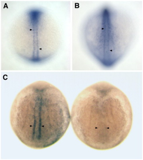
cap1 expression is enriched in adaxial cells prior to and during their pre-migratory behaviors and is dependent on Hedgehog signaling. (A) In a 5-somite stage wild-type embryo, cap1 is expressed specifically in the adaxial cells of both newly formed somites (upper arrowhead) and the presomitic mesoderm (lower arrowhead). (B) At the 12-somite stage, cap1 continues to be expressed within the adaxial cells of somitic and presomitic mesoderm (arrowheads), and becomes increasingly upregulated in the overlying ectoderm. (C) 10-somite stage embryos showing cap1 expression in wild-type (left) and yot mutant (right) embryos. cap1 is not expressed in the adaxial cells of yot mutants (arrowheads), which are deficient in Hedgehog signal transduction. EXPRESSION / LABELING:
|
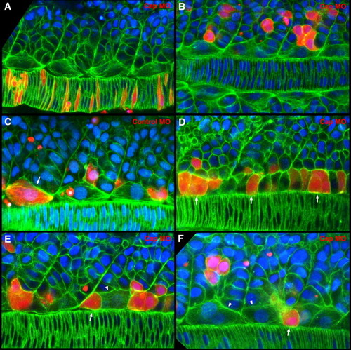
Cap1 is required for adaxial cells to form apical constrictions, intercalate, and broaden. Cells from embryos co-injected with rhodamine-dextran and translation blocking or mismatch control morpholinos were transplanted into presumptive mesodermal precursor regions of wild type embryos at the late blastula stage. Confocal images of adaxial cells at the mid-notochord level of fixed 8–10 somite stage embryos labeled with phalloidin (green) and DAPI (blue). Rhodamine-dextran labels transplanted cells (red). Dorsal view with anterior to the left in all panels. (A, B) Injection of Cap1 morpholino does not cause defects in non-adaxial mesoderm cell behaviors. (A) Cap1 morpholino-containing notochord cells undergo normal intercalation behaviors and are otherwise indistinguishable from wild-type neighbors. (B) Cap1 morpholino-containing cells in the more lateral somite display the same shapes and distributions as their wild type neighbors. (C) Adaxial cells containing mismatch control morpholinos are able to complete the typical array of cell behaviors, becoming broadened and making somite attachments (arrow) in a manner indistinguishable from their wild-type neighbors. (D) Cap1-deficient adaxial cells display the normal cuboidal and columnar cell shapes in presomitic mesoderm (arrows). (E, F) Cap1-deficient adaxial cells fail to make apical constrictions, to intercalate normally, or to broaden and make bipolar somite boundary attachments. Instead they display an isolated rounded appearance (arrows in panels E, F) among neighboring wild-type adaxial cells that have broadened (arrowheads in panels E, F). PHENOTYPE:
|

Cap1-deficient adaxial cells can adopt the slow-twitch myosin-expressing (F59-positive) fate and migrate to appropriate final positions. Shown are three-dimensional reconstructions of confocal images of slow-twitch, F59-positive (green) and Cap1-deficient, rhodamine-dextran-labeled transplanted (red) trunk muscle cells at the 26-somite stage. F59-expressing transplanted cells are yellow. Lateral, dorsal and transverse views of confocal sections containing transplanted cells are shown. Crosshairs highlight the position of the same cell in each view. Anterior is to the right in lateral and dorsal panels. F59-positive, Cap1-deficient transplanted cells can be found in normal slow muscle fiber positions, demonstrating that although Cap1 is required for normal pre-migratory adaxial cell behaviors, it is not required for slow muscle cell fate or for position in the postmigratory, superficial slow fiber (SSF) layer (A) and the non-migratory muscle pioneer (MP) cluster (B) in mosaic embryos. |

Unillustrated author statements EXPRESSION / LABELING:
|
Reprinted from Developmental Biology, 309(2), Daggett, D.F., Domingo, C.R., Currie, P.D., and Amacher, S.L., Control of morphogenetic cell movements in the early zebrafish myotome, 169-179, Copyright (2007) with permission from Elsevier. Full text @ Dev. Biol.