
- Title
-
A role for the Myoblast city homologues Dock1 and Dock5 and the adaptor proteins Crk and Crk-like in zebrafish myoblast fusion
- Authors
- Moore, C.A., Parkin, C.A., Bidet, Y., and Ingham, P.W.
- Source
- Full text @ Development
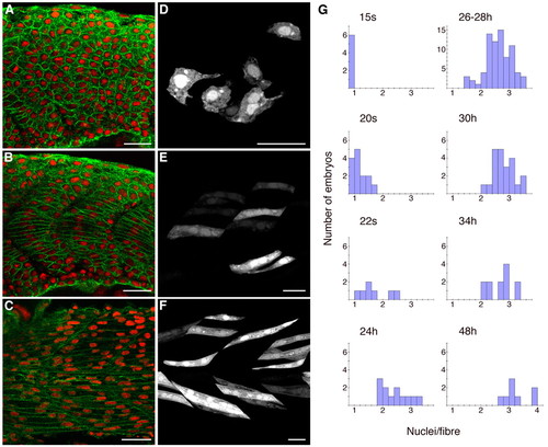
Wild-type fusion of fast-twitch myoblasts. (A-C) Mid-trunk; β-catenin (green) and DAPI (red). (A) At 18 somites; optical section mid-way between the midline and the lateral surface, through fast precursors, showing unfused small rounded cells. (B) At 18 somites; optical section through medial adaxial cells, the slow-fibre precursors, which have elongated. (C) At 26 hpf; optical section through fast muscle fibres, showing multinucleate fused cells. (D-F) mylz2:GFP transgene expression in isolated cells. GFP labelling is found almost exclusively in fast-twitch muscle fibres, the few slow fibres that express the transgene being distinguishable by their shape and more-advanced differentiative state. (D) At 17 somites, showing small rounded mostly mononucleate cells with projections. (E) At 20 somites, showing that fusion has started to occur. Some cells are multinucleate. (F) At 26 hpf, showing elongated multinucleate fibres. (G) Quantitative data showing distributions of mean nuclei/fibre for individual wild-type embryos fixed at progressive embryonic stages, from 15 somites (15s) to 48 hpf (48h). Scale bars: 25 μm. |

Expression of Dock1, Dock5, Crk and Crkl during embryogenesis. (A) Reverse transcriptase (RT)-PCR analysis of dock1, dock5, crk and crkl expression, from the two-cell to 22 somite stage. Two lanes per stage; left-hand lane amplified with five PCR cycles fewer than the right-hand lane. (B-E) In situ hybridisation analysis of dock1 and dock5. (B) dock1, 9 somites (9s). (C) dock1, 22 somites. (D) dock5, 9 somites. (E) dock5, 22 somites. EXPRESSION / LABELING:
|
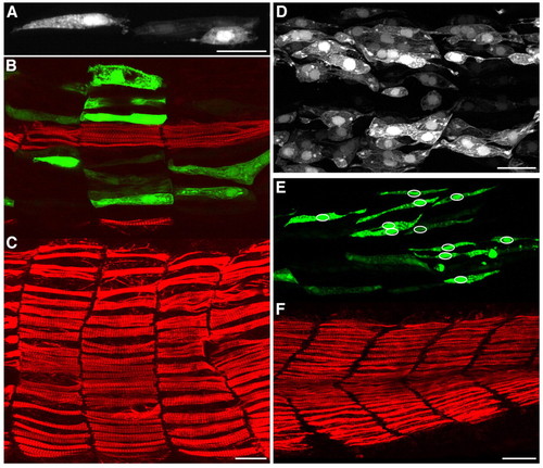
Morphant embryos show defective fast-myoblast fusion. (A) Embryo injected with dock5 splice 1 morpholino, showing examples in neighbouring somites of GFP-positive mononucleate cells that (left) span the somite and (right) have failed to elongate. (B,C) Embryo injected with dock5 splice 2 morpholino, showing an example of defects in somite shape. B is more medial and shows that the GFP-positive cells are medial to the superficial slow fibres shown in C, the slow fibres in B being the medially located muscle pioneers. (D) Embryo injected with crkl ATG morpholino, showing an example with many mononucleate GFP-labelled cells that have failed to elongate and still extend thin processes. (E,F) Embryo injected with dock5 splice 1 and crkl ATG morpholinos combined. Slow fibres are unaffected (F), although there are many mononucleate GFP-positive fast fibres (E). Circles in E indicate the positions of nuclei. All morpholinos were injected in solution containing mylz2:GFP plasmid and were fixed at 26-28 hpf. Green, GFP of mylz2:GFP plasmid; red, F59-antibody slow-fibre stain. Scale bars: 25 μm. PHENOTYPE:
|

Quantitative data showing the distributions of mean nuclei/fibre for individual embryos treated with dock1, dock5, crk and crkl morpholinos. These data are based upon injection of morpholinos at concentrations optimised to produce negligible early embryonic arrest. PHENOTYPE:
|

RT-PCR analysis of splice morpholino function at 26-28 hpf. (A) Changes in size and intensity of the amplification product between wild-type (wt) and MO-injected (MO) embryos indicates effectiveness of splice morpholinos. The dock1 wild-type band decreased in intensity when the embryos were injected with dock1 splice MO, although a 40 bp deletion was detectable by sequencing. dock5 splice 1 morpholino caused inclusion of 273 bp of intron. dock5 splice 2 morpholino caused a 75 bp deletion. crk morpholino caused excision of 578 bp. crkl morpholino mainly caused inclusion of 57 bp of intron, although a deletion of 472 bp was also detected by sequencing. All changes created premature stop codons. (B) Diagrammatic representation of the Dock and Crk protein structures and the truncations produced by the splice morpholinos. Sites recognised by morpholinos are marked with arrows. Affected exon boundaries are shown with red vertical lines. Dock proteins of the DOCK180 subfamily possess two CZH domains (green) and one or two C-terminal proline-rich PXLPXK motifs (*), which bind Crk. The adaptor proteins Crk and Crkl contain an SH2 domain (dark blue) and two SH3 domains (pale blue). The N-terminal SH3 domain binds to DOCK180 proteins. EXPRESSION / LABELING:
|
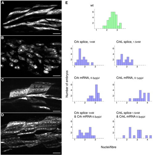
Overexpression of Crk and Crkl causes excess fusion of fast myoblasts by 26-28 hpf, and rescues splice morphants. (A-D) Examples of GFP-positive cells in embryos injected with mylz2:GFP plasmid alone (A) or together with 1.5 mM of crkl splice morpholino (B), 0.1 μg/μl crkl mRNA (C), or 0.3 μg/μl crkl mRNA and 1.5 mM crkl splice morpholino combined (D). (E) Quantitative data showing distributions of mean nuclei/fibre for individual embryos injected with crk and crkl splice morpholinos and/or crk and crkl mRNA. Scale bars: 25 μm. |