
- Title
-
Early developmental specification of the thyroid gland depends on han-expressing surrounding tissue and on FGF signals
- Authors
- Wendl, T., Adzic, D., Schoenebeck, J.J., Scholpp, S., Brand, M., Yelon, D., and Rohr, K.B.
- Source
- Full text @ Development

Thyroid development is impaired in hands off mutant zebrafish embryos. Anterior is to the left. Stages are indicated bottom left, genotype top right and staining/marker bottom right. Arrows show thyroid primordium; arrowheads show pharyngeal endoderm. Ventral (A,B,Q), dorsal (C,D) and lateral (E-P) views are shown. T4 (thyroid hormone) immunostaining (A,B,Q) and in situ hybridisation (C-P). (A,B) In hans6 embryos, no T4-producing follicles are detectable. (C,D) Pharyngeal endoderm appears to be normal in hans6 mutants. (E-K) Expression of thyroid developmental markers. (L-N) The thyroid differentiation marker slc5a5 is expressed in approximately 10% of hans6 mutant embryos. (O-Q) In hanc99 mutants, both primordium and differentiated thyroid are reduced. hy, hypothalamus; mhb midbrain-hindbrain boundary. |

han is expressed in tissues surrounding the thyroid primordium. (A-C) Whole-mount embryos, anterior is up; (D-F) sections. Labelling of panels is as in Fig. 1. (A,B) The thyroid primordium (arrow in B) is adjacent to the outflow tract of the heart (arrows in A). Two expression domains between the first and the second branchial arch (black arrowheads) flank the thyroid on the same anteroposterior level. Red arrowheads point to han (hand2) expression in the fin buds. (C,D) Thyroid hormone (TH) immunostaining visualises arch-associated neurons (AANs) in the putative carotid body primordium (arrowheads). Double staining of hand2 and TH (D) shows that both expression domains are adjacent to each other. (E) hand2 is also expressed in the endoderm (red arrowheads). Notice the strong expression in cells next to AANs (black arrowheads), and in the heart (arrows). (F) For comparison, see the thyroid marker in F (combined with the heart marker MF20). h, heart. EXPRESSION / LABELING:
|
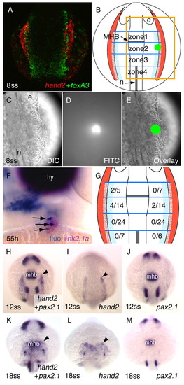
Fate mapping of thyroid precursor cells reveals their close association with the lateral plate mesoderm. (A) Double fluorescence in situ hybridisation of han (hand2; red) and the endoderm marker foxa3 (green). Dorsal view, anterior is up. (B) Subdivision of the region of interest (light blue: potential overlap between lateral plate mesoderm and endoderm) into four zones according to landmarks. Red: lateral plate mesoderm; green, see D,E. The orange square corresponds to the section shown in C-E. (C-E) Example of photoactivation: (C) Nomarski view, (D) after photoactivation, (E) overlay. (F) Example of an embryo (frontal view) in which photoactivated cells (fluorescein, dark blue, arrows) are detectable in the thyroid primordium (pink). Photoactivated cells are also present in pharyngeal cells further away from the midline. (G) Numbers of embryos in which photoactivated cells contributed to the thyroid. Given are numbers of such embryos/total numbers of uncaging experiments at the corresponding anteroposterior level. Compare with B. Notice that only photoactivation in zone 1 and 2 resulted in contributions to the thyroid. (H-M) Comparison of hand2 expression with the midbrain-hindbrain boundary (MHB) marker pax2.1. Arrowheads point to the anterior border of hand2 expression. e, eye; hy, hypothalamus; mhb, midbrain-hindbrain boundary; n, notochord; ss, somite stage. EXPRESSION / LABELING:
|
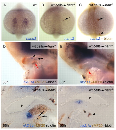
Grafted wild-type cells can restore nk2.1a expression in hans6 mutant embryos. Labelling of panels is as in Fig. 1. Anterior up (A-C) or to the left (D,E), and sections (F,G). (A-C) Grafted wild-type cells express han (hand2) in the mutant background. Arrows point to a single grafted cell that contributed to the anterior lateral plate mesoderm. The arrowhead in C indicates a grafted cell that contributed to other tissue, not expressing hand2. (D-E) Examples of hosts fixed at 55 hours post fertilisation (hpf). D shows an embryo (corresponding to #4 in Table 1) that has restored nk2.1a expression (arrowhead), an occurrence that was never seen in siblings. The black arrow points to grafted wild-type cells, the red arrows to the heart rudiment. E shows a hans6 embryo without restored thyroidal nk2.1a expression. (F,G) Cross sections of specimens of hans6 embryos with restored nk2.1a expression. Grafted cells in the embryo shown in F (corresponding to #5 in Table 1) are adjacent to the thyroid in cartilage, but also in heart tissue (arrows). Note also that such a large thyroid as in F was never observed in untreated hans6 embryos. Often, donor cells clearly belong to the pharyngeal mesenchyme, but are also close to the heart rudiment (G, embryo corresponds to #3 in Table 1). c, cartilage precursor cells; h, heart rudiment; p, pharynx; t, thyroid. |

Elimination of han expression sites in the pharyngeal area does not influence thyroid development. Labelling is as in Fig. 1. (A-D) In edn1 morphants (B), han (hand2) expression in the first (1) and third branchial arches is eliminated. In tfap2a(low, lockjaw) morphants (C), hand2 expression in the second branchial arch (2) is missing. In double morphants (D), hand2-expressing cells are only present in arch-associated cells (arrowheads), as well as some weak expression in the endoderm (confirmed on sections, data not shown). (E) The thyroid (arrow) appears normal in edn1/tfap2a double morphants. (F) In foxi1 morphants, hand2 expression in arch-associated cells (arrowhead) is specifically ablated. (G) Normal expression of nk2.1a in foxi1 morphants (arrow). h, heart. |
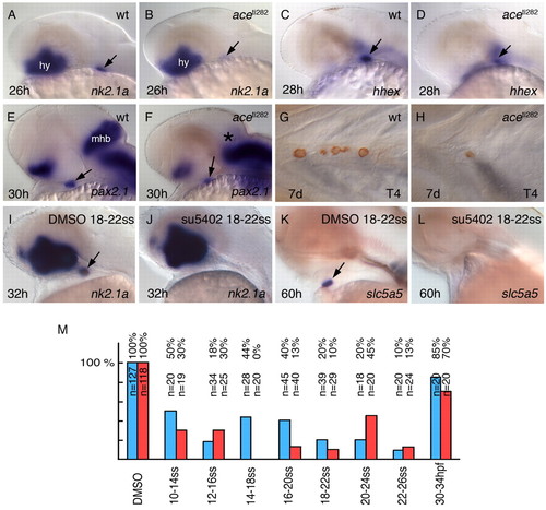
FGF signalling is required for thyroid development. Labelling is as in Fig. 1. Anterior is to the left. Lateral (A-F,I-L) and ventral (G,H) views are shown. (A-F) Expression of thyroid markers in ace mutants and in wild-type (wt) siblings. (G,H) ace larvae have a strong reduction in thyroid gland size. (I,K) In DMSO-treated control embryos, the thyroid appears normal. (J,L,M) Following different stages of su5402 treatment, the thyroid is completely lost. (J,L) Example embryos without thyroid; (M) the complete data set. Blue bars, embryos with thyroidal nk2.1a expression; red bars, slc5a5 expression. Arrows point to the thyroid primordium; the asterisk indicates the absent midbrain-hindbrain boundary (MHB) in ace mutants.hy, hypothalamus; mhb, midbrain-hindbrain boundary, ss somite stage. |

FGFs act downstream of or in parallel to han. Labelling is as in Fig. 1. Grafting of FGF-soaked beads significantly increases the number of hans6 mutant embryos with detectable slc5a5 expression. (A) Example of a hans6 mutant implanted with an FGF1-soaked bead. Implantation of FGF-soaked beads significantly increased the probability that hans6 mutants expressed detectable thyroid markers (arrow). Counterstaining against MF20 enables the identification of hans6 mutant homozygotes. (B) Diagram showing the complete data set. (C) Schematic drawing, summarising scenarios of how FGF signalling and Hand2 might influence thyroid development. han-expressing cells (blue) influence early thyroid development either directly (I,II) or indirectly (III). (I) Hand2-expressing tissue signals directly to the thyroid precursor cells (green) via the FGF pathway (arrow) to promote the development of the precursor cells. (II) Hand2-expressing tissue has a direct influence on thyroid development (not via FGF signalling), with neighbouring tissue providing additional FGF signals. In this scenario, increased levels of FGFs can compensate for the missing influence of Hand2-expressing cells. (III) In the case of indirect influence of Hand2-expressing tissue on thyroid development, FGF signals can be involved at different levels (i.e. on the level of the upper or the lower arrow, or on the level of both arrows). Green cells: thyroid precursor cells; blue cells: Hand2-expressing cells; red arrows: potential contribution of FGF signals. b, bead; h, heart. |

Unillustrated author statements |