
- Title
-
Distinct Functions for Different scl Isoforms in Zebrafish Primitive and Definitive Hematopoiesis
- Authors
- Qian, F., Zhen, F., Xu, J., Huang, M., Li, W., and Wen, Z.
- Source
- Full text @ PLoS Biol.
Identification of Zebrafish scl-β Isoform (A) Northern blot analysis of scl expression in 3-h, 1- to 2-somite (s), 5- to 6-somite, 9- to 11-somite, 18-somite, and 2-dpf embryos and adult kidney. The 3′-probe (3′ UTR) recognized both scl-α and -β isoforms (middle), whereas the 5′-probe (5′ UTR) detected only the full-length scl-α (top). ef1α was used as the loading control (bottom). (B) Gene structures of scl-α and -β. scl-α consists of 4 exons (boxed regions). scl-β transcript is initiated from the middle region of exon 2, and a potential TATA-box sequence (indicated in bold capital letters) is predicted at position -31 of the transcription initiation site. The black bars indicate the positions of scl-α MO, scl-β MO, and scl-sp MO. The white boxes represent sequences that encode the basic helix-loop-helix domain. |

Distinct Expression Pattern of scl-α and -β during Early Embryonic Development (A–L) Examination of scl-α and -β expression by WISH in wild-type and mon mutant embryos. WISH was performed in 2-somite (s) (A and G), 4-somite (B and H), 8-somite (C and I), 18-somite (D and J), and 26-hpf (E and K) wild-type embryos, and 18-somite mon mutant embryos (F and L). The 3′-probe (A–F) detected the expression of both scl-α and -β, whereas the 5′-probe (G–L) detected scl-α expression. The arrows indicate the expression of scl-β (E), but not scl-α (K), in the ventral wall of DA. Embryos are in dorsal (A and G), flat-mounted dorsal (B, C, H, and I), and lateral (D–F and J–L) views with anterior to the top (A and G) or the left (B–F and H–L). (M–R) Transverse sections through the trunk region of 26-hpf wild-type embryos with dorsal up. Fluorescence in situ hybridization showed the expression of c-myb in the ventral wall of DA (white arrowhead in [M, O, P, and R]). Immunochemistry staining showed that Ab-Scl-C (black arrowhead in [N and O]) but not Ab-Scl-N (Q) detected Scl expression in the ventral wall of DA, demonstrating the co-localization of scl-β and c-myb in the ventral wall of DA (O). NC, notochord; NT, neural tube; PCV, posterior cardinal vein. (S) Schematic diagram in flat-mounted dorsal view to illustrate the temporal and spatial expression of scl-α and -β. Red represents scl-β expression, whereas yellow indicates scl-α/β co-expression. EXPRESSION / LABELING:
|
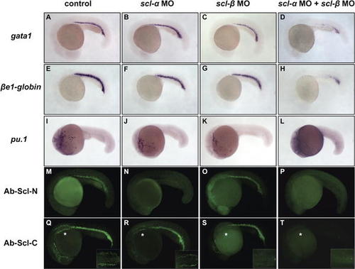
scl-α and -β Are Functionally Redundant in the Initiation of Primitive Hematopoiesis (A–L) WISH of gata1 (A–D), βe1-globin (E–H), and pu.1 (I–L). Similar expression of gata1, βe1-globin, and pu.1 were detected in 20-hpf control embryos (A, E, and I), scl-α morphants (B, F, and J), and scl-β morphants (C, G, and K). In contrast, expression of gata1, βe1-globin, and pu.1 were significantly reduced or absent in scl-α/β co-injected morphants (D, H, and L). All embryos are in lateral view with anterior to the left. (M–T) Immunohistochemistry staining of endogenous Scl proteins in 20-hpf control embryos (M and Q) and scl-α (N and R), scl-β (O and S), and scl-α/β co-injected (P and T) morphants using Ab-Scl-N (M–P) and Ab-Scl-C (Q–T). In scl-α morphants, the Ab-Scl-N (N), but not Ab-Scl-C (R), staining in the ICM was absent, showing that scl-α MO specifically blocks the translation of scl-α. In the scl-β morphants, the Ab-Scl-C staining was selectively abolished in the APLM, where only scl-β is transcribed ([S] asterisk, inset), demonstrating that scl-β MO specifically blocks the protein expression of scl-β. In the scl-α/β co-injected morphants, neither Ab-Scl-N nor Ab-Scl-C staining was detected (P and T). Insets in (Q–T) are dorsal views of the magnified APLM marked by asterisks. Embryos are in lateral view with anterior to the left. EXPRESSION / LABELING:
|
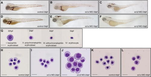
Requirement of scl-α and -β at Different Development Stages of Primitive RBC Differentiation (A–F) O-dianisidine staining of hemoglobin. At 3 dpf, expression of hemoglobin was severely reduced in scl-β morphants (C) compared to control embryos (A) and scl-α morphants (B). At 5 dpf, the o-dianisidine staining in both scl-α (E) and scl-β (F) morphants was dramatically decreased compared to that in control embryos (D). All embryos are in lateral view with anterior to the left. (G–L) May-Grunwald Giemsa staining of RBCs of different developmental stages (30 hpf to 5 dpf). Normal primitive RBCs at 30 hpf to 5 dpf can be classified into four main developmental stages (G): stage I, basophilic erythroblast; stage II, polychromatophilic erythroblast; stage III, orthochromatophilic erythroblast; and stage IV, mature erythrocyte. While RBCs from 2-dpf control embryos (H) and scl-α morphants (I) have differentiated into the polychromatophilic erythroblast stage (stage II), RBCs from scl-β morphants (J) are halted at the earlier basophilic erythroblast stage (stage I). At 4 dpf, RBCs from scl-α morphants (L) are arrested at stage II, while there is normal development of stage IV RBCs in control embryos (K). Scale bar (G–L), 10 μm. |
The scl-α and -β Transcripts in 30-hpf and 2-dpf Embryos and RBCs. Virtual Northern blotting shows that 30-hpf RBCs express both scl-α and -β (lane 2) whereas 2-dpf RBCs express predominantly scl-α (lane 4). |

Specification of Definitive Hematopoietic Stem Cells Requires scl-β but Not scl-α (A–F) WISH of definitive hematopoietic stem cell markers c-myb (A–C) and runx1 (D–F). Expression of c-myb and runx1 in the ventral wall of DA was markedly reduced in 30-hpf scl-β morphants (C and F) but not in 30-hpf control embryos (A and D) or scl-α morphants (B and E). Insets are high-magnification (20×) views of the ventral wall of DA. The arrowheads indicate the definitive hematopoietic stem cells along the ventral wall of DA. (G–I) WISH of T cell marker rag1. rag1 expression in thymic T cells was abolished in 5-dpf scl-β morphants (I) but not in control embryos (G) and scl-α morphants (H). Arrows indicate the thymus region. In all panels, embryos are in lateral view with anterior to the left. EXPRESSION / LABELING:
|
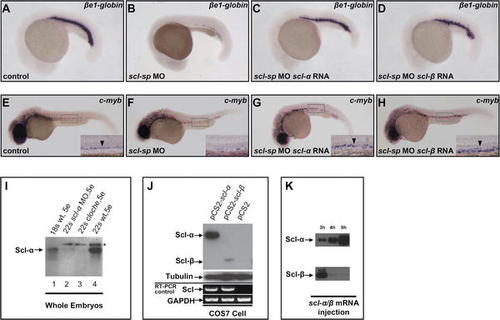
Differences in Protein Expression Levels of Scl-α and -β Confer Their Distinct Functions (A–H) βe1-globin and c-myb expression in scl-sp morphants is rescued by scl-α and -β mRNA. (A–D) show WISH of βe1-globin in 20-hpf control embryos (A), scl-sp morphants (B), scl-sp morphants injected with scl-α mRNA (C), and scl-sp morphants injected with scl-β mRNA (D). (E–H) show WISH of c-myb in 30-hpf control embryos (E), scl-sp morphants (F), scl-sp morphants injected with scl-α mRNA (G), and scl-sp morphants injected with scl-β mRNA (H). Insets in (E–H) are high-magnification (20×) views of the ventral wall of DA, indicated by black arrowhead. In all panels, embryos are in lateral view with anterior to the left. (I) Immunoblotting with Ab-Scl-C antiserum shows the expression levels of Scl-α and Scl-β proteins in wild-type (wt) embryos, clo mutant embryos, and scl-α morphants. Although Scl-β protein was not detectable, immunoblotting of whole embryo protein extracts (50 μg for each sample) showed that Scl-α protein expression increased as embryos developed from the 18-somite (lane 1) to 22-somite (lane 4) stage. The clo mutant embryos (lane 3) and scl-α morphants (lane 2), in which Scl-α protein was not detected, were used as the controls to distinguish the Scl-α protein (arrow) and nonspecific band (asterisk). (J) Immunoblotting with Ab-Scl-C antiserum shows the expression levels of Scl-α and Scl-β proteins in transfected COS7 cells. Western blotting of whole cell protein extracts (10 μg for each sample) prepared from COS7 cells transfected with full-length scl-α, scl-β, or blank vector constructs revealed that the protein level of Scl-α was much higher than that of Scl-β. RT-PCR analysis (bottom) showed similar RNA levels of scl-α and -β in these transfected cells. Tubulin and GAPDH were used as controls for Western blot and PCR, respectively. (K) Immunoblotting with Ab-Scl-C antiserum shows the expression levels of Scl-α and Scl-β proteins in the embryos injected with in vitro synthesized scl-α or -β mRNA. Western blotting of whole embryo protein extracts (50 μg for each sample) from embryos injected with 500 pg of scl-α or -β mRNA showed that, at 3 h post-injection, the Scl-β protein was detected as being at a comparable level to that of Scl-α but dramatically reduced by 4 h post-injection. EXPRESSION / LABELING:
|

The Artery Endothelial Cells Are Retained in Both scl-α and -β Morphants. WISH of artery-specific markers deltaC ([A], n = 42/42; [B], n = 41/44; and [C], n = 45/46) and grl ([D], n = 40/40; [E], n = 45/46; and [F], n = 44/47). At 24 hpf, expression of deltaC and grl was only slightly reduced in both scl-α ([B and E], arrowheads) and -β morphants ([C and F], arrowheads) when compared to the control embryos ([A and D], arrowheads). EXPRESSION / LABELING:
|