
- Title
-
The disarrayed mutation results in cell cycle and neurogenesis defects during retinal development in zebrafish
- Authors
- Baye, L.M., and Link, B.A.
- Source
- Full text @ BMC Dev. Biol.
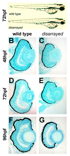
Delayed retinal lamination in disarrayed eyes. (A) Lateral views of wild-type (upper) and disarrayed (lower) sibling embryos at 72 hpf. Mutant embryos are characterized by smaller eyes and forebrain by 42 hpf. Transverse central retinal sections of wild-type (B,D, F) and disarrayed (C,E,G) at 48 hpf (B,C), 72 hpf (D,E), and 96 hpf (F,G). Note the significant delay in differentiation and lamination in the disarrayed retina, although lens growth appears normal. |
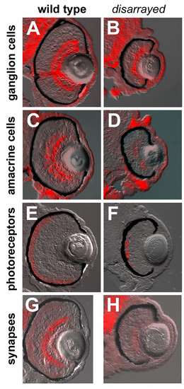
Retinal cell-type marker analysis in disarrayed eyes. Transverse central retinal sections from 96 hpf wild-type (A,C,E,G) and disarrayed (B,D,F,H) embryos assessed for cell-type and lamina specific markers. (A,B) Immunofluorescence for zn8 antigen (retinal ganglion cells and their axons); (C, D) parvalbumin (amacrine cells and their processes); (E,F) zpr1 antigen (cone photoreceptor cells); and (G,H) SV2 antigen (synaptic vesicles). All cell types are present and in the correct laminar position. All figures are bright field images overlaid with the immunofluorescent label. PHENOTYPE:
|

Proliferation defects in disarrayed retinas. (A) One hour labeling with BrdU (red) at 48 hpf (i, ii), 72 hpf (iii, iv) and 96 hpf (v, vi) in wild-type and disarrayed embryos. Wild-type proliferative cells are restricted to the marginal zone by 72 hpf, whereas the disarrayed retinal progenitors do not restrict to the marginal zone until 96 hpf. (B) Quantization of the one hour BrdU-injections presented as the percentage of BrdU-positive cells divided by total cell number at 48 hpf, 72 hpf and 96 hpf. ** p d 0.01 (Student's t-test). Grey bars, wild-type; black bars, disarrayed. Three central retinal sections were counted from three independent embryos for each time point. |
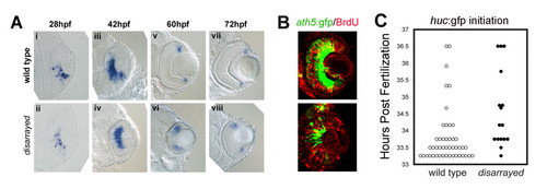
Initiation of retinal neurogenesis in disarrayed eyes. (A) In situ analysis of ath5 expression during retinogenesis in wild-type (i, iii, v, vii) and disarrayed eyes (ii, iv, vi, viii). (B) BrdU pulse-labeling (red) in wild-type (upper) or disarrayed (lower) embryos carrying the ath5:gfp transgene (green). Embryos were injected with BrdU at 36 hpf and fixed at 42 hpf. Note that in both genotypes, ath5:GFP expression in central retinal cells is post-S-phase. (C) Developmental time (hours post fertilization) for the initiation of huc:GFP expression. The initiation of GFP expression was recorded for all embryos at 0.5 hr intervals in a clutch from an in-cross of disarrayed heterozygous parents (one circle represents one embryo; mutant (black circles) and wild-type (white circles)). Note that all embryos initiate huc:GFP expression within the same window of development. Results from one representative experiment (n = 16 mutant and n = 45 wild-type embryos from one clutch of embryos). EXPRESSION / LABELING:
|
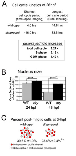
Extended cell cycle and reduced rate of retinal neurogenesis in disarrayed. (A) Cell cycle kinetics at 26 hpf as determined using either direct time-laspse imaging or Cumulative BrdU Labeling with Labeled Mitosis methods. The total cell cycle period as well as S-phase and G2+M-phases were significantly extended at 28 hpf in disarrayed retinas, as indicated by fold increase compared to wild-type siblings. (B) Nucleus size in wild-type (WT, grey) or disarrayed (dry, black) retinal progenitors at 24 and 48 hpf. (C) Comparison of the proportion of proliferative cells (red) to total cells (red + grey) which exited that cell cycle from 28–34 hpf in wild-type and disarrayed embryos. In mutant eyes there are fewer total cells (216.2 +/- 8.4 vs. 383.1 +/- 15.0; n = 15 and n = 14 respectively) and a lower percentage that have exited the cell cycle by 34 hpf (average +/- SE, n = 18 (WT) and n = 11(dry)). ** p d 0.001 (Student′s t-test). |
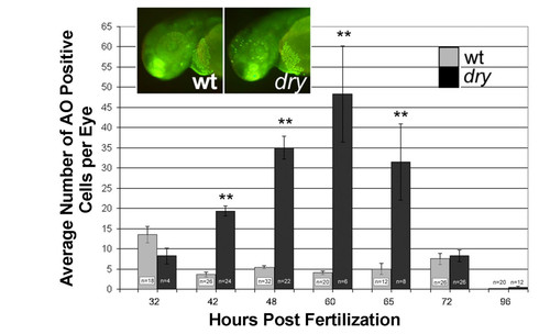
Elevated cell death in disarrayed retinas. Acridine orange (AO) was used to label dying cells in the living zebrafish embryo. The average number of positive cells per retina in wild-type (grey bars) and disarrayed embryos (black bars) is presented. Inset shows chromatin fluorescence (green) in wild-type (left) and disarrayed (right) embryos following acridine orange treatment at 48 hpf. Note that there are significantly more cells labeled in the disarrayed mutant retina and forebrain when compared to wild-type. ** p ≤ 0.01 (Student's t-test). n = total number of embryos counted. PHENOTYPE:
|
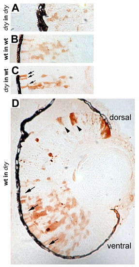
Genetic mosaic analysis of disarrayed retinal cells. Genetic mosaic retinas at 72 hpf where donor cells were lineage-labeled using biotin conjugated dextran (brown). (A) disarrayed mutant cells in a disarrayed mutant host embryo. Note reduced morphological differentiation and lamination of cells. (B) Wild-type cells in a wild-type host embryo. (C) disarrayed mutant cells in a wild-type host embryo. The disarrayed cells show wild-type morphology and lamination in the wild-type environment (compare B and C). For example, morphological rescue of photoreceptor elongation can be seen (C, arrows). (D) Wild-type cells in a disarrayed mutant embryo. Small wild-type cell clones resemble mutant cells as they appear delayed in morphological differentiation and lamination (dorsal, arrowheads). Wild-type cells in clones with greater donor mosaicism have normal morphological differentiation and rescue adjacent disarrayed mutant cells (ventral, arrows). The number of mosaic embryos examined: n = 2 disarrayed into disarrayed, n = 5 wild-type into wild-type, n = 9 disarrayed into wild-type, n = 8 wild-type into disarrayed. Each embryo contained multiple cell clones. |
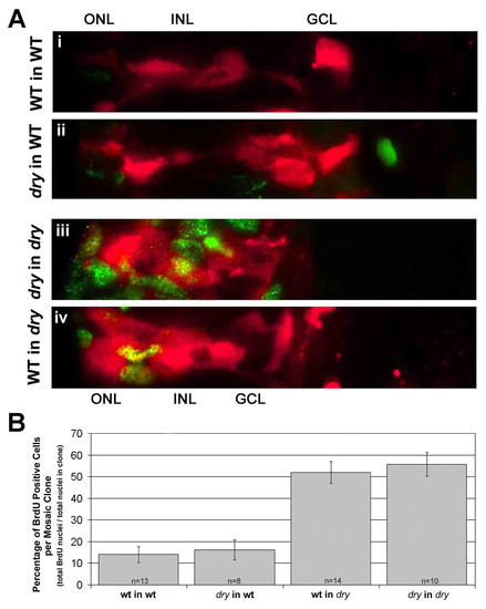
Defects in cell cycle exit for disarrayed are cell-non-autonomous. (A) Genetic mosaic retinas at 61 hpf where donor cells were lineage-labeled using rhodamine conjugated dextran (red). Chimeras were injected with BrdU for one hour at 60 hpf and processed for BrdU detection (green/yellow). Donor cells are shown in the outer nuclear layer (ONL), inner nuclear layer (INL) and ganglion cell layer (GCL) regions. Cells that are double labeled with the linage marker (red) and BrdU (green) appear yellow. (i) Wild-type cells in a wild-type host embryo. (ii)disarrayed cells in a wild-type host embryo. (iii) disarrayed cells in a disarrayed mutant host embryo. (vi) Wild-type cells in a disarrayed embryo. Note the increase in proliferative cells of wild-type or disarrayed genotype in mutant retinas (iii and iv, green/yellow cells). (B) Quantization of the percent of BrdU-positive cells within small (2-20 cells) mosaic clones. n = number of mosaic clones quantitated. |

Unillustrated author statements |