
- Title
-
Muscle-specific expression of the smyd1 gene is controlled by its 5.3-kb promoter and 5'-flanking sequence in zebrafish embryos
- Authors
- Du, S.J., Rotllant, J., and Tan, X.
- Source
- Full text @ Dev. Dyn.
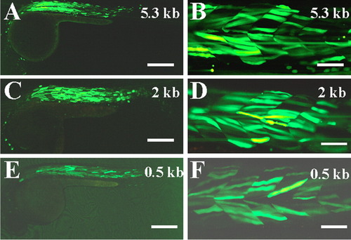
Transient expression analysis of smyd1-gfp in zebrafish embryos. A-F: GFP expression in skeletal muscles of zebrafish embryos injected with smyd1-gfp constructs that contain the 5.3 kb (A,B), 2 kb (C,D), or 0.5 kb (E,F) smyd1 gene promoter and 5'-flanking sequences, respectively. Scale bars = 500 μm in A,C,E, 100 μm in B,D,F. |
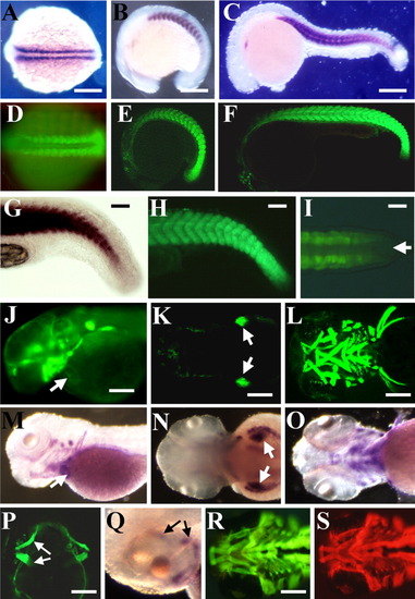
Expression of green fluorescent protein (GFP) reporter gene in developing somites, skeletal, cardiac, head, and fin muscles of transgenic embryos. A-C: In situ hybridization showing expression of smyd1 mRNA in 14 (A), 16 (B), and 22 (C) hours postfertilization (hpf) zebrafish embryos. D-F: GFP expression in developing somites and skeletal muscles of 14 (D), 16 (E), and 22 (F) hpf smyd1-gfp transgenic embryos. G,H: High magnification shows smyd1 mRNA (G) and smyd1-gfp (H) expression in presomitic mesoderm in the tail bud of 24 hpf embryos. I: Dorsal view of GFP expression in presomitic mesoderm but not in the axial mesoderm in the tail bud of smyd1-gfp transgenic embryos at 24 hpf. The arrow indicates axial mesoderm. J: Side view of GFP expression in cardiac muscles of smyd1-gfp transgenic fish at 2 days postfertilization (dpf). K,L: GFP expression in pectoral fin (K, dorsal view) and cranial muscles (L, ventral view) at 2 or 3 dpf, respectively. M-O: In situ hybridization showing expression of smyd1 mRNA in cardiac (M), pectoral fin (N), and cranial muscles (O). Arrows in M and N indicate heart and pectoral fin, respectively. P,Q: Expression of GFP (P, dorsal view) or smyd1 mRNA (Q, side view) in eye muscles of smyd1-gfp transgenic larvae at 5 dpf. R,S: Colocalization of GFP expression and MF20 staining in craniofacial muscles at 4 dpf. GFP expression (R) and MF20 staining (S) were directly observed on the same smyd1-gfp transgenic embryo using a green or a red filter, respectively. Scale bars = 150μm in A-C,R,S, 30μm in G,H, 15 μm in I, 200 μm in M-O,P,Q. EXPRESSION / LABELING:
|

Characterization of E-box mutations on the activity of smyd1 promoter by transient expression analysis in zebrafish embryos. All panels are side views showing green fluorescent protein (GFP) expression in skeletal muscles of zebrafish embryos at 38 hours postfertilization (hpf). From A to H, zebrafish embryos were injected with the following DNA constructs: A: [smyd1-gfp (0.5)]; B: [smyd1-gfp (a*)]; C: [smyd1-gfp (a*b*)]; D: [smyd1-gfp (a*b*c*)]; E: [smyd1-gfp (a*b*c*d*)]; F: [smyd1-gfp (a*b*c*d*e*)], G: [smyd1-gfp (a*b*c*d*e*f*)], and H: [smyd1-gfp (a*b*c*d*e*f*g*)], respectively. Scale bar = 80 μm. |
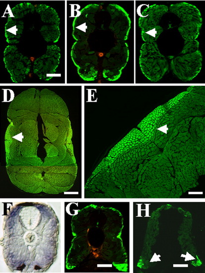
Different levels of green fluorescent protein (GFP) expression in slow and fast muscles of smyd1-gfp transgenic fish. A-C: GFP expression directly photographed on embryonic sections at 4 days postfertilization (dpf) under a confocal microscope. Slow muscles express higher levels of GFP than fast muscles in three transgenic lines of smyd1-gfp transgenic fish. A, line-27; B, line-32; C, line-51. Arrows indicate slow muscles. D,E: Cross-section showing higher levels of GFP expression in slow muscles of line-32 at 2 months old. Slow muscles are indicated by arrows. F: Cross-section shows smyd1 mRNA expression in both slow and fast muscles at 4 dpf, although the staining appeared slightly stronger in superficial slow muscles. G,H: Cross-sections (dorsal on top) showing GFP expressing slow muscles in wild-type transgenic larvae (G) or yot mutant larvae (H) at 4 dpf. Slow muscles are clearly present at the dorsal and ventral myotome in yot mutant embryos. Scale bars = 150 μm in A-C, 500 μm in D, 200 μm in E, 120 μm in G,H. EXPRESSION / LABELING:
|
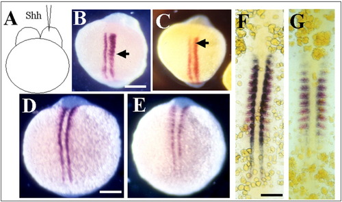
Regulation of smyd1 gene expression in adaxial cells by Hedgehog signal. A-C: Microinjection of Shh mRNA induces smyd1 (B) or myod (C) expression in the injected side as indicated by arrows. D-G: Comparison of smyd1 expression in wild-type (D,F) and smo (E) or yot (G) mutant embryos. smyd1 expression is reduced in the mutant embryos. F and G are dual in situ with smyd1 (blue) and myod (red). Scale bars = 200 μm in A-C, 150 μm in D, 75 μm in E. EXPRESSION / LABELING:
|