
- Title
-
Mutations in laminin alpha 1 result in complex, lens-independent ocular phenotypes in zebrafish
- Authors
- Semina, E.V., Bosenko, D.V., Zinkevich, N.C., Soules, K.A., Hyde, D.R., Vihtelic, T.S., Willer, G.B., Gregg, R.G., and Link, B.A.
- Source
- Full text @ Dev. Biol.

bala69 mutants show anterior segment dysgenesis. (A) Comparison of wild-type (top) and two bala69 mutant (middle and lower) zebrafish embryos at 4 dpf. Irregular pupils (middle and lower) are found in all bala69 mutants, whereas the shortened body axis was present in only some embryos (lower). (B) High magnification of the pupil in wild-type (left) and a bala69 mutant (right). (C) Anterior segment histology of wild-type (top) and bala69 mutant (bottom) eyes from 24 hpf to 72 hpf. |
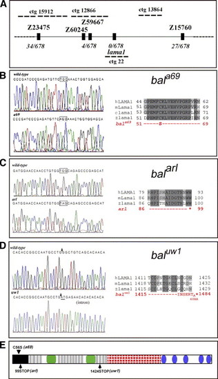
Positional cloning of the bala69 mutation. (A) Genetic map of chromosome 24 containing the bala69 mutation. Microsatellite markers, genomic contigs (ctg), and gene names are indicated with number of recombinants and embryos genotyped. (B–D) Sequence chromatograms showing lama1 mutations in (B) bala69, (C) balarl, and (D) baluw1. For each, DNA sequence for wild-type (top, left) and homozygous mutants (bottom, left) are shown. Altered codons are indicated with boxes and the uw1 splice-site mutation is indicated with arrows. Corresponding protein changes are shown on the right of each panel. Mutant Lama1 sequence (red) is aligned with wild-type sequence from human (h), mouse (m), and zebrafish (z). Light grey highlights show amino acids conserved in multiple species, whereas dark grey highlights indicate residues conserved in all species. Asterisk indicates the termination codon. INSERT^60AA indicates the translated intronic regions predicted from the baluw1 splice-site mutation. (E) Schematic of Lama1 protein with a69, arl, and uw1 mutation positions indicated by arrows. Lama1 domains are indicated: black box—N-terminal domain; light grey rectangles—EGF repeats; green rounded rectangles—laminin B domains; red cross-hatched rectangle—laminin coiled-coil domain; blue ellipses—C-terminal globular domains. |

Laminin alpha 1 expression during eye development. In situ hybridization for lama1 transcripts (purple) was investigated at (A) 24 hpf, (B) 48 hpf, and (C) 72 hpf. Laminin-1 immunoreactivity (red) was examined at (D) 24 hpf, (E) 48 hpf, and (F) 72 hpf and found to be associated with the cornea, sclera, lens capsule, outer segments of photoreceptors, and optic nerve (arrows in panel E). By 72 hpf, immunoreactivity was also observed in the developing anterior segment angle (arrowheads in panel F). EXPRESSION / LABELING:
|

Lens removal and comparison to bala69 mutants. Views of a 24 hpf wild-type embryo (A) before and (B) after surgical lens removal. The intact lens following removal is indicated by the arrowhead. Histology of 10 dpf eyes from (C) wild-type, (D) lens-ablated, and (E, F) two bala69 mutants to show the phenotypic range. |

Anterior segment dysgenesis and reduced focal adhesions. Histology and marker comparisons of 10 dpf wild-type, lens-ablated and bala69 mutant larvae reveal lens-independent ocular phenotypes. (A) Iridocorneal angle shows normal periocular cell differentiation in wild-type (left) and lens-ablated (middle) larvae. Although pigment cell differentiation in bala69 mutants (right) occurs, many cells appear hyperplastic (arrows) and the collagen rich stromal layer (arrowheads) is disorganized. (B) Development of the ciliary canal (red arrows) is initiated normally in wild-type (left), lens-ablated (middle), and bala69 mutant (right) larvae. (C) Cornea differentiation in wild-type (WT), lens-ablated (LA), and two bala69 mutants (bala69). Analysis showed both focal (left mutant) and more severe (right mutant) corneal dysplasias. (D) Immunoreactivity for activated phospho[Y397] FAK in 54 hpf wild-type eyes. Inset d shows a higher magnification of the boxed differentiating lens epithelium and cornea. (E) Immunoreactivity for activated phospho[Y397] FAK in 54 hpf bala69 mutant eyes. Inset e shows a higher magnification of the boxed dysgenic lens epithelium and cornea. Note the robust activated FAK staining in the plexiform layers of both wild-type and mutant eyes, but the lack of intense and punctate staining the neural retina margins, lens cells, and corneal endothelium in the bala69 eyes. (F) Immunoreactivity for Paxillin in 54 hpf wild-type eyes. Inset f shows a higher magnification of the boxed differentiating lens epithelium and cornea. (G) Immunoreactivity for Paxillin in 54 hpf bala69 mutant eyes. Inset g shows a higher magnification of the boxed dysgenic lens epithelium and cornea. Paxillin immunoreactivity, although strong in the ciliary margin, is diffuse and reduced in the cornea. EXPRESSION / LABELING:
|

Retinal ganglion cell and photoreceptor defects with loss of Lama1. Brn3c-GFP expression (A–C, green) in retinal ganglion cells from (A) wild-type, (B) lens-ablated, and (C) lama1 morphant eyes. Note the axon routing mistakes in lama1 morphants, but not within lens-ablated eyes. Zpr-1 immunoreactivity (D–F, red) shows photoreceptors in retina from (D) wild-type, (E) lens-ablated, and (F) bala69 mutants. Note the ectopic, mis-positioned photoreceptors in the mutants (arrowheads), but not in lens-ablated eyes. |

lama1 morphants show hyaloid vascular defects. Transgenic fli1:gfp zebrafish were used to assess hyaloid vascular development in (A) wild-type, (B) lens-ablated, and (C, D) two lama1 morphants. Images are projected confocal z-stacks from 50 μm series. |

Ocular genetic mosaics of bala69 mutant and wild-type cells. (A) 24 hpf genetic mosaic embryos showing a high contribution of lineage-labeled donor cells (red) to ocular structures. (B) 5 dpf whole embryo phenotypes of wild-type donor to wild-type host (top) and bala69-donor to wild-type host ocular chimeras (bottom). Note the irregular pupil in the absence of other body phenotypes in the bala69 to wild-type ocular mosaic. (C) Ocular phenotype of wild-type to wild-type mosaics (top) show no abnormalities, whereas (D, E) bala69 to wild-type ocular mosaics show a spectrum of phenotypes including (D) pinhole pupils and severe microphthalmia and (E) iridophore patterning defects with cataracts. |

Histological comparison of adult wild-type mosaic, lens-ablated, and bala69 mosaic eyes. (A–D) Iridocorneal angle analysis in (A) wild-type mosaics showed normal morphology including annular ligament and ciliary canal. In (B) lens-ablated fish, cell differentiation in the angle appeared normal, although the ciliary canal was often narrow. Angle differentiation in (C, D) bala69 mutant mosaics was often abnormal including defects in the annular ligament (arrows), iris, and ciliary margin. In the posterior region, the iridophore layer (arrowheads) appeared normal in (E) wild-type mosaics and (F) lens-ablated fish but was often thick in (G) bala69 mutant mosaics. Whereas the retinal ganglion cell (RGC) layer was dysmorphic in both lens-ablated and bala69 mutant mosaics with lens degeneration, focal dysplasias within the RGC layer were also observed in (I) bala69 mutant mosaics with normal lenses. No dysplasias or other phenotypes were noted in (H) wild-type mosaics. |
Reprinted from Developmental Biology, 299(1), Semina, E.V., Bosenko, D.V., Zinkevich, N.C., Soules, K.A., Hyde, D.R., Vihtelic, T.S., Willer, G.B., Gregg, R.G., and Link, B.A., Mutations in laminin alpha 1 result in complex, lens-independent ocular phenotypes in zebrafish, 63-77, Copyright (2006) with permission from Elsevier. Full text @ Dev. Biol.