
- Title
-
Knockdown of Nav1.6a Na+ channels affects zebrafish motoneuron development
- Authors
- Pineda, R.H., Svoboda, K.R., Wright, M.A., Taylor, A.D., Novak, A.E., Gamse, J.T., Eisen, J.S., and Ribera, A.B.
- Source
- Full text @ Development

Two primary spinal neuron types, RB cells and CaPs, expressed scn8a. (A) Hu immunoreactivity revealed RB somata in a 48 hpf embryo. (B) In situ hybridization indicated that dorsal spinal cord neurons contained scn8a transcripts. (C) Dual examination of Hu immunoreactivity and scn8a mRNA identified RB cells as the dorsal neurons that express scn8a. (D) GFP+ neurons (arrowheads) in Tg(flh:GFP) embryos projected their axons (arrows) to ventral muscle, indicating that they were CaPs. In addition, GFP+ neurons were ISL+ (arrowheads, yellow nuclei), as were RB cells (asterisks). In two of the three hemisegments shown, VaPs were present with adjacent CaPs. VaPs are essentially duplicated CaPs that do not extend their axons as far ventrally, because of competition (Eisen, 1992; Eisen and Melancon, 2001). (E) GFP+ neurons (arrowheads) in Tg(flh:GFP) embryos did not express isl1 mRNA (carats), a marker of MiP. In the middle hemisegment, both CaP and VaP were present. (F) GFP+ CaPs (arrowheads) in Tg(flh:GFP) embryos expressed scn8a, as did dorsal RB cells (asterisks). (G) Double in situ hybridization for isl2, a marker of CaP, and for scn8a revealed that both CaPs (arrowheads) as well as RB cells (asterisks) expressed scn8a. (A-C) Dorsal views, (D-G) lateral views, anterior is towards the left and dorsal is upwards. Scale bars: in C, 30 μm for A-C; in G, 30 μm in D-F; 15 μm in G. EXPRESSION / LABELING:
|
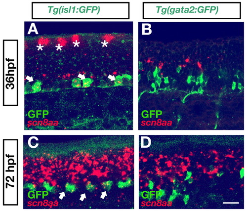
Dorsally projecting, but not ventrally projecting, SMNs expressed scn8a mRNA. GFP+ dorsally projecting SMNs in Tg(isl1:GFP) embryos expressed scn8a mRNA at 36 (A) and 72 (C) hpf (arrows). Asterisk in A indicates RB cells. (B) By contrast, ventrally projecting SMNs [GFP+ in Tg(gata2:GFP) embryos] did not express scn8a at either 36 (B) or 72 (D) hpf. Scale bar: 25 μm in A,C; 40 μm in B,D. EXPRESSION / LABELING:
|
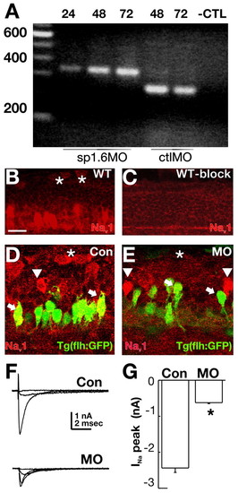
Injection of 1.6MO blocked splicing and abolished Na+ channel immunoreactivity and current in spinal cord neurons that expressed scn8a mRNA. (A) In RNA isolated from embryos injected with the splice blocking MO, excision of the 96 bp intron between exons 3 and 4 did not occur. This was true at 24, 48 and 72 hpf. By contrast, embryos injected with a control splice MO displayed normal splicing at this junction. (B-E) Na+ channel immunoreactivity in control and morphant embryos. (B) A pan-specific Na+ channel antibody revealed immunoreactivity in several classes of spinal cord neurons in 48 hpf wild-type embryos. RB cells (asterisks) and ventral neurons were brightly labeled. (C) Preincubation of the antibody with peptide blocked immunodetection of Na+ channels. (D) GFP+ ventral neurons (arrowhead, arrows) in Tg(flh:GFP) embryos displayed Na+ channel immunoreactivity. (E) The 1.6MO reduced Na+ channel immunoreactivity in some (arrows, asterisks) but not all (arrowheads) spinal neurons. Scale bar: 10 μm. (F) Na+ currents recorded from RB cells of control embryos had larger amplitudes than those recorded from 1.6MO morphants. (G) Peak Na+ current amplitude was significantly reduced in RB cells of 1.6MO morphants (n=6) versus control (n=6) embryos (*P≤0.001). EXPRESSION / LABELING:
|

RB cells survived longer in 1.6MO morphants embryos. (A) Injection of the 1.6MO prolonged RB survival in 72 hpf 1.6MO morphants embryos. RB cells were revealed by Hu immunoreactivity. Dorsal views are shown. Scale bar: 25 μm. (B) At 72 hpf, there were 40-fold more RB cells in 1.6MO morphants (n=12) versus control (n=9) embryos (*P≤0.003). EXPRESSION / LABELING:
PHENOTYPE:
|
Ventrally projecting motor axons displayed abnormal trajectories in 1.6MO morphants even though CaP axons project normally. (A-D) Motor axon morphology was assayed using SV-2 immunoreactivity (green) in conjunction with a marker of the muscle postsynaptic receptor, α-bungarotoxin (red). At 48 hpf (A,B), little difference was noted in znp-1 immunoreactivity of motor axons of 1.6MO morphants (B) compared with control-injected embryos (A). However, by 72 hpf, there were marked differences (C,D). Motor axons of 1.6MO morphants (D, asterisks) branched more than those of control-injected embryos. Moreover, in morphants (D), there was a reduction in the normal alignment between motor axons and postsynaptic receptors, assessed by the near absence of green and abundance of yellow in C versus the reduction in yellow and increase in green in panel D. (E-L) Injection of 1.6MO did not affect axon outgrowth from CaP when synapses first form. (E-H) At 21-22 hpf, znp-1 immunoreactivity revealed no differences between ventrally projecting CaP axons in either control (E,F) or 1.6MO-injected (G,H) embryos. Embryos were squash-mounted and, consequently, motor nerves on both sides of an embryo were often present in a single confocal section (e.g. F,H). (I,J) CaPs were directly labeled by dye injection (green) at 20 hpf and embryos were fixed at 21-22 hpf and stained for SV-2 immunoreactivity. Injection of either ConMO (I) or 1.6MO (J) did not affect CaP axon morphology. (K,L) In addition to axonal morphology (SV-2, green), postsynaptic specializations, as detected by α-bungarotoxin labeling (red) were not altered by injection of either Con (K) or 1.6 (L) MO. Scale bars: in A, 20 μm for A,B; 30 μm for C; in E, 50 μm for E-H; in J, 10 μm for I,J; in L, 20 μm for K,L. PHENOTYPE:
|

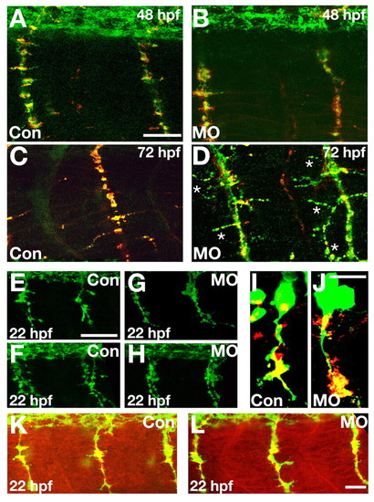
Injection of 1.6MO altered SMN axons although Mauthner axons projected normally. (A-F) Zn-8 immunoreactivity revealed differences in SMN axon outgrowth between morphants and controls. Between 48 and 66 hpf (A-D), innervation of the dorsal musculature was reduced in 1.6MO morphants (B,D) versus control (A,C). Arrows and arrowheads in C,D,F indicate the rostral and dorsal projections. Asterisks in C indicate ventral projections. At 78 hpf (E,F), substantial recovery had occurred in 1.6MO morphants (F) and the majority of dorsal somites were innervated. (G) Although 1.6MO morphants (squares) initially displayed no innervation of the dorsal musculature, this phenotype recovered. Recovery began at 3 dpf and was nearly complete by 5 dpf. Circles represent data from control embryos. (H) In ventral muscle at 66 hpf, extra ventrally projecting axons (asterisks) were present in 28.8±3.6% of segments in 1.6MO morphants but were not present in controls. (I,J) At 78 hpf, the axons of ventrally projecting SMNs in 1.6MO morphants branched more (J) compared with controls (I), consistent with results obtained with the znp-1 antibody (cf. Fig. 5C,D). (K-N) Injection of 1.6MO did not affect Mauthner cell axonal morphology. (K,L) Mauthner cell axons projected caudally into spinal cord in 48 hpf 1.6MO morphants (L) and controls (K). (M,N) Mauthner axons of 72 hpf 1.6MO morphants (N) displayed morphologies similar to those of controls (M). Scale bars: in F, 40 μm for A-F; in H, 40 μm for H and 20 μm for I,J; in L 40 μm for K,L; in N, 40 μm for M,N. EXPRESSION / LABELING:
PHENOTYPE:
|

Injection of 1.6MO but not ConMO perturbed outgrowth of SMN axons in Tg(gata2:GFP) embryos. (A-E) GFP+ ventrally projecting axons displayed more branching in Tg(gata2:GFP) morphant (B,D,E) but not control (A,C) embryos at 72 hpf. Embryos were squash-mounted and, consequently, motor nerves on both sides of an embryo were often present in a single confocal section (e.g. A,B). Scale bars: in B, 100 μm for A,B; in E, 50 μm for C-E. EXPRESSION / LABELING:
|

Effects of the 1.6MO on ventrally projecting SMN axons are non cell-autonomous. In wild-type embryo mosaics (A,B), the presence of the 1.6MO (B) or ConMO (A) in ventrally projecting SMNs did not produce abnormally projecting axons. The lineage tracer fluorescein-conjugated dextran (green) was co-injected with the MO. SMN axons were revealed by zn8 immunocytochemistry (red). Ventrally projecting axons (arrows) are both zn8+ (red) and GFP+ (green), indicating that they contain axons of neurons derived from the MO-injected cell. (C,D) In embryos of the Tg(gata2:GFP) line, GFP+ (green) axons were abnormal even though they did not contain the lineage tracer rhodamine-conjugated dextran (red). However, other spinal neurons were positive for the lineage tracer (red), indicating that the 1.6MO had non cell-autonomous effects of ventrally projecting SMNs. Each panel contains a projection of six consecutive confocal sections. Scale bar: 40 μm. EXPRESSION / LABELING:
PHENOTYPE:
|