
- Title
-
Prdm1 acts downstream of a sequential RA, Wnt and Fgf signaling cascade during zebrafish forelimb induction
- Authors
- Mercader, N., Fischer, S., and Neumann, C.J.
- Source
- Full text @ Development
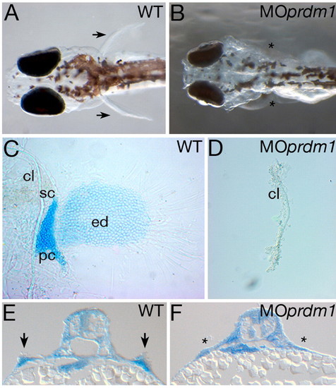
prdm1 morphants lack pectoral fins. (A,B) Dorsal views of wild-type (WT, A) and prdm1 morphant (MO prdm1, B) four-day-old larvae. C,D) Cartilage stainings of wild-type and prdm1 morphant pectoral fins at four days postfertilization. Note that prdm1 morphants only develop a cleithrum. (E,F) Methylene Blue-stained transverse cryosections of 48 hpf embryos. Arrows point towards pectoral fins in the wild type and asterisks indicate the absence of pectoral fins in the morphant. cl, cleithrum; ed, endochondral disc; sc, scapulocoracoid; pc, postcoracoid process. |

Expression of prdm1 compared with fgf24 and tbx5 during limb bud initiation. (A-C) prdm1 whole-mount in situ hybridization at embryonic stages prior to limb bud initiation. Lateral views of a 12-somite (A) and a 15-somite (B) stage embryo revealing prdm1 (blue) and myod (red) expression. Note prdm1 expression overlapping with myod in the somites. (C) Lateral view of a 17-somite stage embryo. Arrows in A-C reveal the most anterior limit of prdm1 expression within the lateral plate mesoderm. (D-F) Dorsal views of 18-somite stage embryos hybridized with prdm1 (D), fgf24 (E) or tbx5+myod (F) riboprobes. Arrows in D point towards the onset of prdm1 expression in the pectoral fin primordia. Note that the prdm1 and fgf24 expression domains are very similar, and that the tbx5 expression domain is broader than the prdm1 domain. (G-I) Dorsal views of 23-somite stage embryos hybridized with prdm1 (G), fgf24 (H) or tbx5+myod (I) riboprobes. Arrows in G point towards the expanded prdm1 expression domain in the pectoral fin. EXPRESSION / LABELING:
|

Prdm1 acts downstream of retinoic acid and Wnt2b signaling during limb bud initiation. (A,B) Lateral views of the retinaldehyde dehydrogenase 2 (raldh2) expression pattern in 12-somite stage wild-type (A) or prdm1 MO-injected (B) embryos. Anterior is to the left. A' and B' depict close-up views of lateral plate mesoderm (arrows) and somite expression at the axial level of pectoral fin formation. (C,D) Expression of the retinoic acid degrading enzyme cyp26a1 in wild-type and prdm1 morphant embryos at the 12-somite stage. Arrow indicates cyp26a1 expression within somites 1 and 2. Note that there is no difference in raldh2 and cyp26a1 expression between prdm1 morphants and wild types. (E-F') prdm1 in situ hybridization of control DMSO-treated 17-somite stage embryos (E,E') and DEAB-treated embryos (F,F'). Note that DEAB treatment leads to a reduction of prdm1 expression within branchial arches (arrow in E) and anterior somites (compare straight and dotted lines in E' and F'). (G,H) prdm1 in situ hybridization on 24 hpf wild-type (G) and neckless mutant (H) pectoral fin bud regions. Arrows in G indicate fin buds; asterisks in F and H indicate a absence of prdm1 expression within the corresponding structure. (I-L) Expression pattern of wnt2b mRNA in wild-type (I), prdm1 morphant (J), 16 µM SU5402-treated (K) and neckless mutant (L) embryos at 24 hpf. Panels show dorsal views with anterior to the top. Note that wnt2b expression is normal in MOprdm1 and SU5402-treated embryos, but is lost in the raldh2 mutant nls (asterisks). DEAB, diethylaminobenzoic acid; DMSO, dimethylsulfoxide; nls, neckless; MOprdm1, prdm1 morphant; WT, wild type. EXPRESSION / LABELING:
|
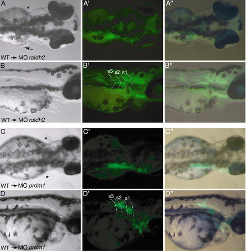
Mosaic analysis in raldh2 and prdm1 morphants. (A) Dorsal view of a three-day-old raldh2 morphant embryo revealing rescued pectoral fin outgrowth on the right side (arrow). (A') Dark-field image of the same embryo, showing transplanted GFP-positive cells labeled in green. (A'') Merged bright-field and dark-field images showing green wild-type cells localizing to the anterior somite region. (B-B'') Lateral views of the same MOraldh2 mosaic embryo as in A. Dotted lines in B' indicate somite boundaries. Note strong GFP-expression in somites 1 to 3. (C-D'') Dorsal and lateral views of an MOprdm1 embryo, where transplanted wild-type cells contribute to anterior somites but do not rescue fin outgrowth. (C'',D'') Merged bright and dark field pictures showing GFP-positive wild-type cells incorporated into the left fin. Dotted lines in D' indicate somite boundaries. Asterisks mark the missing pectoral fin. s, somite. |

Expression of fgf24 and tbx5 in MOprdm1-injected embryos compared with ika mutants. (A-H) Dorsal views of fgf24 whole-mount in situ hybridization on wild-type, prdm1 morphant or ikarus mutant embryos, as indicated in each panel. Anterior is to the top. Fgf24 expression is reduced at 18 hpf (B) and is not visible in pectoral fin regions of 20 hpf (D) or 24 hpf (G) MOprdm1-injected embryos. Although the onset of fgf24 expression is normal in fgf24 mutant ikarus embryos (A,E), its expression is not maintained at later stages (H). Asterisks indicate missing fgf24 expression. (I-N) Dorsal views of tbx5 in situ hybridizations on 20 hpf (I-K) and 24 hpf (L-N) wild-type, ika and MOprdm1 embryos. At 20 hpf, no difference can be detected between wild-type, MOprdm1 and ika embryos. At 24 hpf, tbx5 expression is strongly reduced in MOprdm1 and ika fin buds. ika, ikarus; MOprdm1, prdm1 morphant embryo; WT, wild type. EXPRESSION / LABELING:
|

prdm1 expression in dae, ika and hst mutant embryos. (A-M) Dorsal views of prdm1 expression in pectoral fin buds at 20.5 hpf (A-D), 24 hpf (E-H), 30 hpf (I-K) and 36 hpf (L,M) of wild-type, hst, ika and dae mutants embryos, as indicated in each panel. In dae embryos, activation of prdm1 expression is normal (B,F,J) but its expression disappears at 36 hpf (asterisks, M). In ika, weak prdm1 staining can be detected within the fin field at 24 hpf (G). This expression disappears in 30 hpf embryos (asterisks, K). In hst, prdm1 cannot be detected at any stage analysed (marked by asterisks in D and H). dae, daedalu; hst, heartstrings; i ka, ikarus; WT, wild type. EXPRESSION / LABELING:
|

Analysis of cell death in ika mutant pectoral fin mesenchyme. (A-C) TUNEL staining on 30 hpf wild-type (WT, A) and ikarus (ika) mutant (B,C) fin level cryosections. Anterior is to the top. No apoptotic cells could be detected in the fin mesenchyme of ika mutants (n=16, B). In one specimen, a few apoptotic cells could be detected in the apical fin ectoderm but not in the fin mesenchyme (C). Arrows indicate fin bud; asterisks indicate the absence of bud formation in ika mutants. |

Comparison of fgf10, pea3 and bmp2 expression in MOprdm1-injected embryos, and in hst and ika mutants. (A-L) Dorsal views of 30 hpf embryos stained for fgf10 (A-D), pea3 (E-H) or bmp2b (I-L) expression. While fgf10 expression is absent in ika, hst and MOprdm1 embryos (B,C,D), pea3 and bmp2b are transiently expressed in ika (F,J) but not hst or MOprdm1 embryos (G,H,K,L). Asterisks indicate a lack of marker gene expression within the pectoral fin mesenchyme. hst, heartstrings; ika, ikarus; MOprdm1, prdm1 morphant embryo; WT, wild type. EXPRESSION / LABELING:
|

Effect of early Fgf inhibitor treatment on erm, prdm1, fgf24 and tbx5 expression. Whole-mount in situ hybridization on 20.5 hpf (23-somite stage) (A,B,E,F,I,J,M,N) and 24 hpf (C,D,G,H,K,L,O,P) control DMSO and SU5402-treated embryos. Probes are as indicated in each panel. (A-D) erm expression is abolished in SU5402-treated embryos. (E-H) prdm1 expression is downregulated in SU5402-treated embryos. (I-L) Upon SU5402 treatment, Fgf24 is still present at 20.5 hpf but becomes strongly reduced at 24 hpf. (M-P) tbx5 expression is not affected at the 23-somite stage (M,N) but is reduced in 24 hpf embryos upon SU5402 treatment (O,P). EXPRESSION / LABELING:
|