
- Title
-
{alpha}A-crystallin expression prevents {gamma}-crystallin insolubility and cataract formation in the zebrafish cloche mutant lens
- Authors
- Goishi, K., Shimizu, A., Najarro, G., Watanabe, S., Rogers, R., Zon, L.I., and Klagsbrun, M.
- Source
- Full text @ Development

Crystallin proteins levels are diminished in cloche embryos. (A) 2D-gel electrophoresis: Total 2.5 dpf extracts as described in the Materials and methods were analyzed. A spot (20 kDa, pI of 8.8) was identified that was strongly reduced in cloche compared with wild-type (WT) embryos. Spot intensities were analyzed by an image analysis program (ImageJ) and the values were normalized to wild type (100%). (B) The spot was cut out of the gel and identified by MS/MS spectrometry sequencing to be a member of the γ-crystallin family. This protein, designated as embryonic γ-crystallin M2 type1 (EM2-1) was 78% homologous to a recently described zebrafish γ-crystallin M2b (M2b) that had been cloned from adult lens tissue (Wistow et al., 2005). The two sequences were aligned. Identical amino acids are in red. The Reverse Position Specific BLAST program showed two ß/γ-crystallin domains that are underlined in black. (C) γ-crystallin gene expression was analyzed by in situ hybridization at 2.5 dpf. Both lateral and dorsal views are shown. γ-crystallin is expressed solely in the lens. EXPRESSION / LABELING:
|
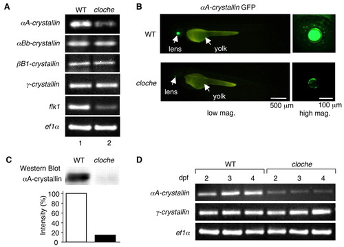
crystallin gene expression. (A) Zebrafish embryo (2.5 dpf) mRNA expression levels were analyzed by semi-quantitative RT-PCR. Lane 1, wild type; lane 2, cloche. Wild-type versus cloche gene expression is shown for αA-crystallin, αBb-crystallin, ßB1-crystallin, γ-crystallin and flk1 (vegfr2). ef1a was used as a loading control. (B) &alphaA-crystallin promoter-EGFP was injected into one- to four-cell stage embryos and fluorescence (green) was measured at 2.5 dpf. Lateral views are shown at low and high magnifications. Fluorescence was found only in the lens and was greatly diminished in the cloche lens. (C) Western blot. Total protein as described in Fig. 1A were extracted and lysates (2 µg) were separated by SDS-PAGE and analyzed by western blotting with anti-αA-crystallin. The bands were analyzed by an image analysis program (ImageJ) and the values were normalized to wild-type bands being 100%. (D) Crystallin expression as function of development. Zebrafish embryo (2-4 dpf) mRNA expression levels were analyzed by semi-quantitative RT-PCR. Wild-type versus cloche gene expression is shown for αA-crystallin and γ-crystallin expression. ef1a was used as a loading control. |

The cloche lens has cataracts. (A) The morphology of the eye was analyzed by bright-field microscopy at 2.5 dpf. (B) Sections were prepared from 2-4 dpf embryos and analyzed by Hematoxylin and Eosin. The green arrow (3 dpf) and red arrow (4 dpf) point to nuclei. (C) The number of lens fiber cell nuclei per section was analyzed at 2-4 dpf. Three sections of each lens were analyzed by an image analysis program (ImageJ). Fourteen embryos for each time point were analyzed. The mean±s.d. is shown. (D) The reflected light (reflectance) of the lens was detected by confocal microscope at 2-4 dpf. The red arrows point to areas of reflected light. (E) Statistical analysis of reflectance intensity. The Mann-Whitney U test was used to test for differences between two groups. The red line in the box corresponds to the median value. The differences between wild-type and clochem39 at 3 dpf (P=0.0339) and 4 dpf (P=0.0209) are significant. (F) The reflected light (reflectance) of the lens in clochem39 (clom39) and cloches5 (clos5) was compared by confocal microscopy at 4 dpf. Reflectance is comparable between the two. The red arrows indicate areas of reflected light. (G) Statistical analysis of reflectance intensity. The Mann-Whitney U test was used to test for differences between two groups. The red line in the box corresponds to the median value. The differences between wild type and clochem39 (clom39) (P=0.0209) or cloches5 (clos5) (P=0.0143) are significant. |
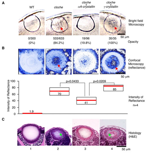
αA-crystallin expression prevents cataract formation in cloche. αA- (panel 3) or γ-crystallin (panel 4) mRNA was injected into embryos at the one- to four-cell stage and phenotypes were compared with uninjected wild type (panel 1) and cloche lens (panel 2). (A) Living embryos were analyzed by stereomicroscopy and the number of opaque lenses was counted. Red arrows point to scattered light. The percent of lenses that demonstrate opacity is shown in parentheses. (B) Living embryos lenses were analyzed by confocal microscopy and the intensity of reflected light was measured. Red arrows indicate areas of reflected light. The median reflectance was measured as in Fig. 3D. The Mann-Whitney U test was used to test for differences between two groups. The differences in the lens between cloche embryos and cloche embryos overexpressing αA-crystallin (P=0.0433) and the differences between cloche overexpressing αA-crystallin versus γ-crystallin (P=0.0209) are significant. (C) Sections were prepared from 4 dpf embryos and analyzed by Hematoxylin and Eosin staining. The green arrows indicate nuclei. The number of lens fiber cell nuclei per section was analyzed at 4 dpf. Three sections of each lens were analyzed by an image analysis program (ImageJ). Fourteen embryos were analyzed. At 4 dpf, wild type and cloche had 0.0±0.0 and 22.6±5.0 nuclei, respectively. In the rescue experiment, αA-crystallin overexpression reduced the nuclei from 22.6±5.0 to 3.0±0.0 (mean±s.d.). |
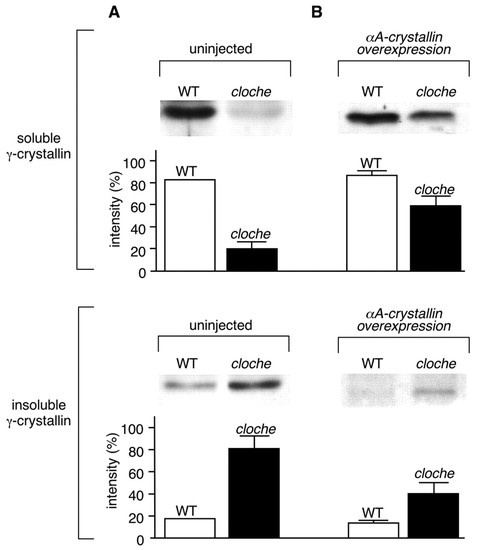
αA-crystallin overexpression prevents *gamma;-crystallin insolubility. (A) Uninjected embryos. (B) Embryos injected with αA-crystallin mRNA. Zebrafish embryos (2.5 dpf) were lysed detergent-free in 20 mM Tris-HCl (pH 7.5) and centrifuged. Soluble (top) and insoluble (bottom) fractions were separated, and were each analyzed by western blot with anti-γ-crystallin antibody. The intensity of the bands in the western blots was measured by an image analysis program. Three independent experiments using clochem39 were performed. The bar graph represents the mean±s.d. |

Retina photoreceptor. Sections were prepared from 3.5 and 5 dpf embryos, and analyzed by immunohistochemistry with antibodies that detect photoreceptors (green). Photoreceptor and retinal thickness are the same in cloche as in wild type. In addition, the photoreceptor structure in cloche does not appear to change from 3.5 to 5 dpf. |

crystallin insolubility and rescue in cloches5. (A) Western blot: total clochem39 and cloches5 embryo extracts were analyzed by western blotting with anti-αA-crystallin antibody. (B) Zebrafish embryo (2-4 dpf) mRNA levels were analyzed by semi-quantitative RT-PCR. Wild-type versus cloches5 embryo gene expression is shown for αA-crystallin and γ-crystallin. ef1a was used as a loading control. (C) αA-crystallin overexpression prevents γ-crystallin insolubility. Zebrafish embryos (2.5 dpf) were lysed detergent-free in 20 mM Tris-HCl, pH 7.5, and centrifuged. Soluble (top) and insoluble (bottom) fractions were separated, and were each analyzed by western blot with anti-γ-crystallin antibody. Two independent experiments using cloches5 were performed. |

Analysis of lens in crash&burn mutants. crash&burn are mutants in bmyb that are developmentally delayed. There were no cataracts (n=80) in these mutants, 2-4 dpf. Thus, developmental delay is insufficient to induce cataract formation. |