
- Title
-
Loss of function of def selectively up-regulates {Delta}113p53 expression to arrest expansion growth of digestive organs in zebrafish
- Authors
- Chen, J., Ruan, H., Ng, S.M., Gao, C., Soo, H.M., Wu, W., Zhang, Z., Wen, Z., Lane, D.P., and Peng, J.
- Source
- Full text @ Genes & Dev.

The defhi429 mutant exhibits hypoplastic digestive organs. (A,B) At 5.5 dpf, the defhi429 mutant had an unabsorbed york sac and exhibited smaller liver and swimbladder and much thinner intestine as seen by dissection microscopes. The liver is outlined with green, the gut with red, the swimbladder with purple, and the york sac with yellow. (mu) Mutant; (wt) wild-type. (C,D) At 5 dpf, Phenol Red injection revealed that the defhi429 mutant had a smaller gallbladder (g) and underexpanded anterior intestine (in). The gut from anterior to posterior (left to right) is outlined with a black line. (E,F) At 5 dpf, alcian blue staining showed that the branchial arches 2-7 in the defhi429 mutant (mu) were deformed. Branchial arches 1-7 in wild type are indicated in E. (G-J) Histochemical staining of alkaline phosphatase showed that pronephric ducts (G,H) at 3 dpf and blood vessels (I,J) at 4 dpf in wild type (wt) and the defhi429 mutant (mu) were indistinguishable. (bv) Blood vessels; (pd) pronephric ducts. EXPRESSION / LABELING:
|

def is required for the expansion growth of intestine, liver, and exocrine pancreas but not endocrine pancreas. (A,B)At2 dpf, whole-mount in situ hybridization using the pan-endoderm marker foxa1 showed that there were no discernible phenotypic differences between wild type (wt) and defhi429 mutant (mu). (in) Intestine; (lv) liver; (p) pancreas. (C,D) At 5 dpf, cross-sectioning showed that the columnar epithelium did form in the mutant gut (mu) but did not fold properly obviously due to greatly reduced cell number when compared with the wild type (wt). (E-H) The expression of gut-specific markers ifabp at 4 dpf (E,F) and alkaline phosphatase at 5 dpf (G,H, yellow arrows) was greatly reduced in the defhi429 mutant. (mu) Mutant; (sw) swimbladder; (wt) wild type. (I,J) At 2 dpf, whole-mount in situ hybridization using a pdx1 probe showed that the initiation and formation of pancreatic bud (p) was not affected in the defhi429 mutant (mu). (in) Intestine. (K,L) At 3 dpf, whole-mount in situ hybridization using a trypsin probe showed that the defhi429 mutant exhibited a much smaller exocrine pancreas. (M,N) In contrast, at 4 dpf, whole-mount in situ hybridization using an insulin probe showed that the endocrine pancreas had no discernible difference between the defhi429 mutant (mu) and the wild type (wt). (O,P) At 4.5 dpf, sectioning in situ hybridization using Fast Red (Roche)-labeled trypsin (for exocrine pancreas, in red) and DIG-labeled insulin (for endocrine pancreas, in purple) probes further confirmed that, when compared with the wild type (wt), the mass of the exocrine pancreas (ex) was greatly reduced while the endocrine pancreas (en) appeared normal in the defhi429 mutant (mu). The mutant epithelium (ep) was narrower and only weakly expressed ifabp (in purple). The liver (lv) in the wild type was labeled using a transferrin probe (for liver, red). (Q,R) At 2 dpf, whole-mount in situ hybridization using prox1 did not reveal a discernible difference between the defhi429 mutant (mu) and the wild type (wt). (S,T) At 4 dpf, the liver bud was obviously smaller and failed to form two lobes in the defhi429 mutant. (U,V) At 5 dpf, cross-sectioning through the esophagus (es) revealed that bile ducts and blood vessels were properly formed in the defhi429 mutant liver (mu) as in the wild-type liver (wt). |
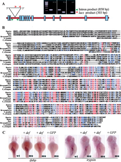
Viral vector insertion in the def gene caused the defhi429 mutant phenotype. (A) Schematic drawing shows the genomic DNA structure of the def gene. The viral vector was inserted in the second intron. The positions of two pairs of primers used for genotyping the mutant embryos are indicated, and their PCR products are shown in the DNA gel photos, respectively. Red arrowheads represent the primer pair for checking the viral insertion in heterozygous (+/-) or homozygous (-/-) mutants; green arrowheads for checking the wild-type gene in wild-type sibling (+/+) or heterozygous mutant (+/-). (B) Amino acid sequence alignment of Def (gi37046654) and its homologs/orthologs in human (gi7657019), mouse (gi20340985), Drosophila (gi20130323), C. elegans (gi17555844), and yeast (gi19076047). (C) Injection of def mRNA (def) rescued the defhi429 mutant phenotype, whereas injection of GFP mRNA (GFP) or mutant defstop mRNA (data not shown) failed to rescue the mutant phenotype. Embryos 3 and 4 d post-injection were used for whole-mount in situ hybridization with ifabp and trypsin probes, respectively. (mu) Mutant; (wt) wild-type. EXPRESSION / LABELING:
|

def encodes a nuclear protein, and its expression is enriched in the endoderm organs. (A, top panel, lanes 2-7) RNA gel blot hybridization revealed that def had two transcribed forms that peaked at 12 hpf and 1 dpf and then gradually reduced to a lower level at 5 dpf. (Lane 9) The shorter form was more abundant in the adult liver (AL). (Lane 8) def expression is undetectable in the defhi429 mutant. (elf1a) elongation factor 1a. (B) Immunostaining using anti-Def antibody showed that Def is a nuclear-localized protein. (C-F) Whole-mount in situ hybridization using a def riboprobe to examine the expression patterns of def at 2 dpf (C), 3 dpf (D), 4 dpf (E), and 5 dpf (F). def expression was specifically enriched in the digestive organs from 3 to 5 dpf. (in) Intestine; (lv) liver; (p) pancreas; (sw) swimbladder. (G,H) Sectioning in situ hybridization showed that def expression is specifically enriched in the intestine (in), liver (lv), and exocrine pancreas (ex) but is excluded from the endocrine pancreas (en) at 3 and 4 dpf. EXPRESSION / LABELING:
|
Loss of function of def selectively up-regulates the expression of EXPRESSION / LABELING:
|
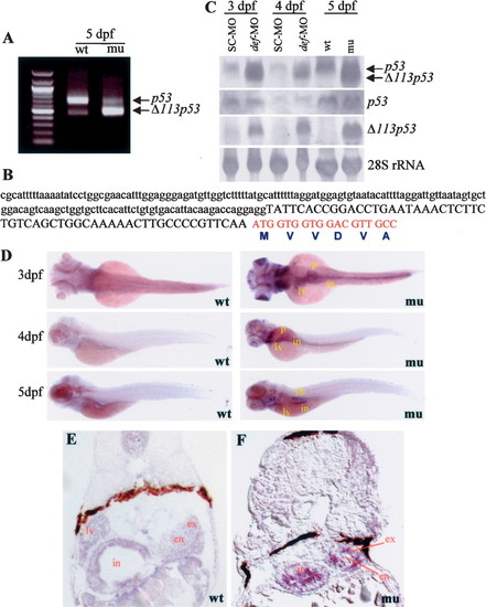
 113p53 in the defhi429 mutant. (A) 5'-RACE reaction identified two p53 isoforms. The longer isoform corresponds to the wild-type p53 (p53) and was predominant in the wild-type embryos. The short isoform (
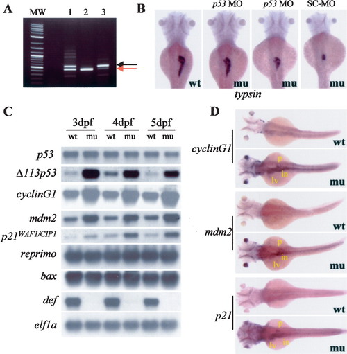
Cell proliferation-related but not proapoptotic-related p53 response genes are up-regulated in the defhi429 mutant. (A) RT-PCR analysis revealed that the p53 MO created an aberrant product with a larger size resulting from insertion of 89 bp from the intron 5. This 89-bp insertion introduced an early stop codon in the p53 mRNA. (MW) Molecular size marker; (lane 1) 0.5 mM p53 MO; (lane 2) standard control MO (SC-MO, human β-globin antisense morpholinos); (lane 3) 1.0 mM p53 MO; (red arrow) wild type p53; (black arrow) misspliced p53. (B) Injection of p53-MOspl targeting p53 mRNA rescued the mutant phenotype. Embryos 70 h post-injection were used for whole-mount in situ hybridization using a trypsin probe. (mu) Mutant; (wt) wild type. (C) Semiquantitative RT-PCR examination of expression of various p53 response genes in defhi429 mutant (mu) and the wild-type control (wt) at 3, 4, and 5 dpf, respectively. Homozygous defhi429 mutants (confirmed by lacking expression of def) had elevated levels of Δ113p53, cyclin G1, p21WAF1/CIP1, and mdm2 but unchanged levels of p53, reprimo, and bax when compared with the wild type (wt). (D) At 3 dpf, whole-mount in situ hybridization using cyclin G1, mdm2, and p21WAF1/CIP-1 probes showed that the expression of these genes was significantly and specifically increased in the digestive organs. (in) Intestine; (lv) liver; (p) pancreas. |

The defhi429 mutant phenotype is caused by compromised cell proliferation. (A,C) BrdU labeling revealed that, at 4 dpf, the number and ratio of cells entering the G1-to-S-phase transition was significantly reduced in the digestive organs of the defhi429 mutant (mu) when compared with the wild type (wt). (Top panel) Cross-sectioning through liver (lv) and intestine (in). (Bottom panel) Cross-sectioning through pancreas and intestine (in). The BrdU incorporation ratios shown in C were obtained by counting BrdU-labeled cells versus total cells in a specific organ (e.g., liver) in sections from seven wild-type and seven mutant embryos, respectively. (en) Endocrine pancreas; (ex) exocrine pancreas. (B,D) At 3 and 4 dpf, histochemical staining using anti-phosphorylated histone 3 (anti-P-H3) revealed that the number and ratio of cells entering G2 to M phase was drastically reduced in the digestive organs of the defhi429 mutant (mu) when compared with the wild type (wt). The ratios of P-H3-positive cells shown in D were obtained by counting P-H3-positive cells versus total cells in a specific organ (e.g., liver) in sections from three wild-type and three mutant embryos, respectively. |
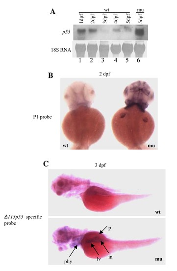
Expression of {delta}113p53 is selectively up-regulated in the defhi429 mutant. (A) RNA gel blot hybridization showed that p53 expression in the wild type peaked at 1 dpf and then gradually reduced to a lower level at 5 dpf (lanes 1 to 5). On the other hand, at 5 dpf, a short p53 isoform ({delta}113p53) (red arrow) rather than the wild type p53 (black arrow) expressed at a very high level in the defhi429 mutant (mu) (lane 6). (B) In situ hybridization using P1 probe as in Fig. 5D showed elevated expression of {delta}113p53 in the defhi429 mutant phenocopies the expression pattern displayed by def (Fig. 4C) at 2 dpf. wt: wild type; mu: mutant. (C) In situ hybridization using an {delta}113p53 specific probe (269 bp) derived from intron 4 (forward primer: 5′-CAGTGGAGGTTGAACATGTCT-3′; reverse primer: 5′-CCTCCTGGTCTTGTAATGTCAC-3′) containing the transcribed 143 bp in {delta}113p53 showed that the expression of {delta}113p53 is specifically enriched in the digestive organs in the defhi429 mutant. wt: wild type; mu: mutant; in: intestine tube; lv: liver; p: pancreas; phy: pharyngeal. |
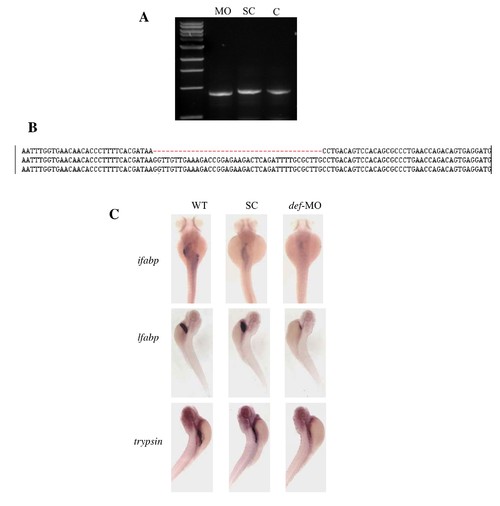
def-MO caused hypoplastic digestive organs as that observed in the defhi429 mutant. (A,B) def-MO (5′-ATGAATATAATGACTTACCAAGCGC-3′) was designed to target the splice junction between exon 2 and intron 2. RT-PCR analysis showed that def-MO created an aberrant frame-shift transcript (A) due to 40 bp deletion in the exon 2 (B). (MO) def-MO; (SC) standard control morpholinos; (C) wild type contol. (C) def-MO morphants were examined with intestinal marker ifabp, liver marker lfabp and pancreas marker trypsin, respectively. 19/27 (70.4%) morphants showed hypoplastic intestine tube (ifabp); 35/47 (74.5%) for liver (lfabp), and 50/56 (89.3%) for pancreas (trypsin). |

At 4.5 dpf, TUNEL assay showed that defhi429 mutant (mu) did not exhibit elevated apoptotic activities when compared to the wild type (wt). White arrow heads mark the apoptotic cells that were mainly from epidermis (mu and wt) and swimbladder (wt). |