
- Title
-
Characterization of two new zebrafish members of the hedgehog family: Atypical expression of a zebrafish indian hedgehog gene in skeletal elements of both endochondral and dermal origins
- Authors
- Avaron, F., Hoffman, L., Guay, D., and Akimenko, M.A.
- Source
- Full text @ Dev. Dyn.
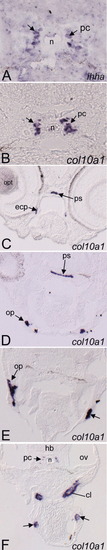
ihha and col10a1 expression in rostro-caudal transverse sections of the head regions of 4dpf zebrafish larvae. A-F: In situ hybridization with an anti sense RNA probe corresponding to ihha (A) and col10a1 (B-F). A: Earliest expression of ihha detected in the skeleton is located in chondrocytes of the parachordal cartilage (pc), which also express col10a1 (B). The latter is more widely expressed, and transcripts were detected in forming dermal bones (C-F). Parasphenoid (ps), ectopterygoid (ecp), opercle (op), cleithrum (cl), and two bilateral clusters of cells, which are part of the pectoral girdle (arrows on F). ect, ectopterygoid; hb, hindbrain; n, notochord; opv, optic vesicle; ov, otic vesicle; pc, parachordal cartilage; ph, pharynx; ps, parasphenoid. EXPRESSION / LABELING:
|

ihha and col10a1 expression in the head skeleton of 6dpf larvae. In situ hybridizations using ihha (A, B, D, G, J, M) and col10a1 (E, H, K, N) probes and Alcian blue staining of cartilage cells (C, F, I, L, O) performed on 12-μm longitudinal (A-C) or transverse (D-O) cryosections of 6dpf larvae. A-C: Longitudinal sections. ihha is expressed in chondrocytes of several cartilaginous elements, such as the Meckel's (me, in A), ceratohyal (ch in B), and parachordal (pc, in B) cartilages. D-O: Cross-sections comparing ihha (left column), col10a1 (middle column) expression, and Alcian blue staining (right column) at different rostro-caudal levels (sections progress caudally). ihha and col10a1 are expressed in chondrocytes (black arrows) of the ceratohyal (ch), hyosymplectic (hs), and parachordal (pc) cartilages. Comparison with Alcian blue staining (right) positively identifies these cells as chondrocytes. In addition to cartilage cells, col10a1 is also expressed in flat cells surrounding the cartilage and several forming dermal bones (black arrowheads). White arrowheads on G, H, M, and N show chondrocytes expressing neither ihha nor col10a1, suggesting that they are less differentiated. See text for details. at, atrium; ba, branchial arches; bp, basal plate; bs, basibranchial; ch, ceratohyal; ep, ethmoid plate; ect, ectopterygoid; fb, forebrain; g, gut; hb, hindbrain; hs, hyosymplectic; me, Meckel's cartilage; n, notochord; op, opercle; opv, optic vesicle; ov, otic vesicle; pc, parachordal cartilage; ph, pharynx; ps, parasphenoid; pq, palatoquadrate; tc, trabecula. EXPRESSION / LABELING:
|
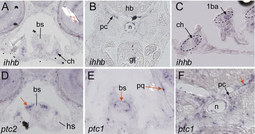
ihhb, ptc1, and ptc2 expression in 6dpf larvae. A-G: In situ hybridizations conducted on transverse sections of 6dpf zebrafish larvae submitted to ISH using a ihhb (A-C), ptc2 (D), or ptc1 (E,F) probes. A-C: ihhb expression is detected in ceratohyal (ch in A), parachordal (pc in B), and branchial arches (1ba in C) chondrocytes, and is similar to ihha. D,F: The pan-hedgehog receptors ptc1 and ptc2 (red arrows) are expressed in the periphery of chondrocytes of several cartilaginous structures, which express ihha or ihhb. In the parachordal cartilage (pc in F), ptc1 expressing cells co-localize with hypertrophic chondrocytes, which also express ihha (Fig. 4M), col10a1 (Fig 4N), and ihhb (B). 1ba, First branchial arch; bs, basibranchial; ch, ceratohyal; hb, hindbrain; g, gut; n, notochord; pc, parachordal cartilage; pq, palatoquadrate. EXPRESSION / LABELING:
|
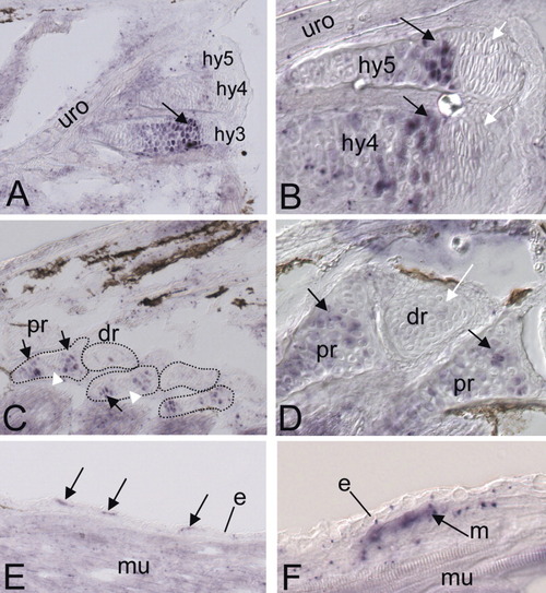
ihha expression in fin endoskeleton and scales. A-F: In situ hybridization using a ihha probe on longitudinal sections of 10-mm larvae. A,B: Caudal fin endoskeleton. A: ihha expression in the hypural of the caudal fin. B: A closer view of A reveals that the distalmost chondrocytes of the hypurals (white arrows) present a different morphology, and do not express ihha. C,D: Dorsal fin endoskeleton. C: ihha is expressed in cells located at the extremities of the proximal radial (pr, black arrows), but not in central cells (white arrowhead). D: Closer view of the dorsal fin endoskeleton showing ihha expression in chondrocytes of the proximal radials (black arrows), but not in the distal radials (white arrows). E: ihha is expressed in growing scales (arrows). Higher magnification (F) shows that ihha expression is undetectable in the epidermis and appears to be restricted in the underlying mesenchyme (arrow on F). e, epidermis; dr, distal radials; hy, hypurals; m, mesenchyme; mu, muscle; pr, proximal radials; uro, urostyle. All sections are oriented anterior to the left and dorsal to the top. EXPRESSION / LABELING:
|
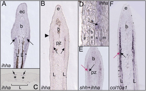
Expression of ihha and col10a1 during fin ray regeneration. In situ hybridization with probes corresponding to ihha (A-D), both ihha and shh (E) and col10a1 (F) on section of zebrafish caudal fin regenerates at 2- and 4-days post amputation (dpa). A,B, D-F: Longitudinal sections; C: transverse section. A: ihha expression is first detected in two faint domains in the blastema compartment (b) at the tip of each amputated hemiray (L), which may correspond to differentiating bone matrix-secreting cells, or scleroblasts. B-D: At 4dpa, during the regenerative outgrowth, ihha-expressing cells are found in two rows of cells adjacent to the basal layer of the epidermis (bel, in D). C: Transverse section of the regenerate at the level of the arrowhead indicated on B. Each hemiray region (L) contains two discrete ihha expression domains (arrows), which signal an imminent bifurcation event and labels the presumptive sister rays (*). Closer view of the epidermis-mesenchyme interface (D) confirms that ihha-expressing cells correspond to scleroblasts (arrow). E: Double in situ with shh and ihha shows that the shh domain (red arrow), which is restricted to the basal epidermal layer, is facing ihha-positive cells (black arrow). F: col10a1 is strongly expressed in both epithelial cells (red arrow) and scleroblasts, from the patterning zone to the proximal part of the regenerate. All longitudinal sections are oriented distal part to the top. bel, basal epidermal layer; e, epidermis; ec, epidermal cap; l, lepidotrichia; m, blastema mesenchyme; pz, patterning zone; *, presumptive sister rays. EXPRESSION / LABELING:
|

Unillustrated author statements |