
- Title
-
Mutation in the delta-subunit of the nAChR suppresses the muscle defects caused by lack of Dystrophin
- Authors
- Etard, C., Behra, M., Ertzer, R., Fischer, N., Jesuthasan, S., Blader, P., Geisler, R., and Strähle, U.
- Source
- Full text @ Dev. Dyn.

Structure of myofibrils in sopfixe mutants. A,B: Birefringence at the flank of wild-type (A) and sopfixe (B) embryos. C-J: Wild-type (C,E,G,I) and sopfixe (D,F,H,J) embryos stained with antibodies against slow muscle myosin (C-F), troponin T (G,H), alpha-actinin (I,J) at 26 hours postfertilization (hpf; C-H) or 48 hpf (I,J). Immunoreactivity of slow muscle myosin highlights the A band (E, F) and α-actinin marks the Z-disc. The I band and M band are indicated by an asterisk and arrow, respectively. E,F: The length of the A band (a = 1.63 μm for wild-type [wt], E, and 1.3 μm for sopfixe, F) and the width of the myofibril (b = 2.3 μm for wt, E, and 1.9 μm for sopfixe, F) are reduced in the mutant. Schematic representation of the sarcomere structure in wild-type and sopfixe embryos. Note the enlargement of the M band and the shortening of the A band. Lateral views, anterior to the left. Scale bars = 100 μm in A,B; 10 μm in C,E; 2.5 μm in E,F; 15 μm in G,H; 8 μm in I,J. |

sopfixe mutants lack clustered acetylcholine receptors. A,B: The 72 hours postfertilization (hpf) embryos were stained with Alexa-green-conjugated α-bungarotoxin. Clusters of acetylcholine receptor (AChR) were not detected in sopfixe mutants (B) but are present in wild-type embryos (A). C,D: Wild-type (A) and sopfixe mutant (B) embryos stained with anti-acetylated tubulin antibody at 72 hpf. Axons of primary motor neurons are present in sopfixe (D) embryos as in wild-type embryos. E: Fluorescein isothiocyanate (FITC) -dextran marked wild-type muscle cell in a sopfixe mutant environment shows clustered acetylcholine receptors (arrows) at 24 hpf. Lateral views, anterior to the left, dorsal up. Scale bars = 30 μm in A-D; 12 μm in E. |
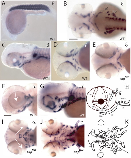
Expression of acetylcholine receptor (AChR) subunits in wild-type and sopfixe fish. A-E: Expression of the δ-subunit in wild-type (A-D) and sopfixe mutant (E) embryos. The δ-subunit is expressed exclusively in muscles. No differences in expression levels between wild-type and sopfixe mutants were detected (compare D with E; A, 24 hpf; B-E, 50 hpf). F,G,I,J: Comparison of the expression of the α1-subunit (F,I) and the γ-subunit (G,J) at 72 hpf. In contrast to all other subunits analyzed, the α1-subunit does not continue to be expressed in eye muscles after 50 hpf (compare F, I, arrow, with G,J). H,K: Schematic drawing of the muscles in the head in lateral (H) and ventral (K) views. ah, adductor hyoideus; am, adductor mandibulae; ao, adductor operculi; do, dilator operculi; hh, hyohyoideus; hp, hypaxial muscles; ih, interhyoideus; ima, intramandibular anterior; imp, intramandibular posterior; io, inferior oblique; ir, inferior rectus; lap, levator arcus palatini; lr, lateral rectus; mr, medial rectus; pf, pectoral fin; sh, sternohyoideus; so, superior oblique; tv, transversus ventralis. A,C,F,G,H: Lateral view; B: dorsal view; D,E,I,J,K: ventral view. Scale bars = 80 μm in A, 60 μm in B-E, 50 μm in F-J. EXPRESSION / LABELING:
|
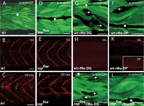
sopfixe suppresses the muscle defects in embryos lacking Dystrophin. A-C: Uninjected wild-type embryos showing expression of the α-actin:GFP transgene (A) or expression of dystroglycan (DG, B) or the combination of Dystrophin (DP) immunofluorescence and α-bungarotoxin staining (C). D-F: sopfixe mutants showing expression of the α-actin:GFP transgene (D) or expression of DG (E) or the combination of DP immunofluorescence and α-bungarotoxin staining (F). Lack of α-bungarotoxin in sopfixe does not affect the pattern of DG or DP expression (compare B,C with E,F). G,H: Wild-type embryos injected with the morpholino against DG (Mo-DG). G: Removal of DG causes detachment of the α-actin:gfp labeled myofibrils from the vertical myosepta (arrows). H: The injected Mo-DG completely abolishes DG expression. I: sopfixe embryo injected with Mo-DG shows a similar pattern of muscle defects as wild-type embryos, in which DG was knocked down, indicating that sopfixe does not suppress the muscle phenotype in Mo-DG morphants. J,K: Embryos in which DP was knocked down show detachment of myofibrils (J) and also a reduction in DG as well as DP staining at the vertical myosepta (K, and insert in K, respectively). L: sopfixe mutant embryos in which DP was knocked down do not show detachment of myofibrils from the vertical myoseptum. Thus, sopfixe suppresses the muscle defects in Mo-DP morphants. A,B, D,E,G-L: 72 hours postfertilization (hpf); C,F: 24 hpf embryos. Asterisk, horizontal myosepta. GFP, green fluorescent protein. Scale bars = 30 μm in A,B,D,E,G-L; 25 μm in C,F. |

Unillustrated author statements EXPRESSION / LABELING:
|