
- Title
-
HSPG synthesis by zebrafish Ext2 and Extl3 is required for Fgf10 signalling during limb development
- Authors
- Norton, W.H., Ledin, J., Grandel, H., and Neumann, C.J.
- Source
- Full text @ Development

Phenotype of daedalus mutant embryos. (A,B) Dorsal views of live 5-day-old sibling (A) and dae mutant (B) larvae, anterior towards the left. dae mutants lack pectoral fins, but appear otherwise normal. (C,D) Alcian Blue staining of the pectoral fin endoskeleton. Wild-type endoskeleton (C) consists of a pectoral girdle, postcoracoid process and endoskeletal disk attached to the cleithrum. dae mutants (D) retain only a dysmorphic pectoral girdle and cleithrum. (E,F) Transverse cryosections of 40 hpf mutant and sibling embryos stained with Methylene Blue. Mutant embryos (F) have a smaller undifferentiated fin bud, without an apical ridge (ar, arrow), when compared with siblings (E). ed, endoskeletal disc; pp, postcoracoid process; sc, scapulocoracoid. |

Positional cloning of the dae locus. (A) SSLP analysis places the dae mutation between two novel SSLP markers, scaf1747_9 (6/810 meioses) and scaf1747_7 (2/698 meioses) on linkage group 21. The fgf10 gene was subsequently identified as a candidate for dae. (B) The tbvbo allele has a K to stop mutation in amino acid 5, and the t24030 allele has an M to V mutation within amino acid 170 of Fgf10. (C,D) Injection of an Fgf10 morpholino directed against the exon2/intron2 splice acceptor site [e2i2 MO (red bar in B)] into wild-type embryos phenocopies the dae mutation. Injection of 0.125 mM morpholino (D) causes a severe truncation of the pectoral fin, identical to the phenotype seen in dae. (E) PCR amplification of the fgf10 open reading frame demonstrates splicing defects following morpholino injection (primer positions indicated in B). L, ladder; –, negative control; +, positive control; c, uninjected (compare with C); d, MO injected, i.e. dae-like phenotype (compare with D). |

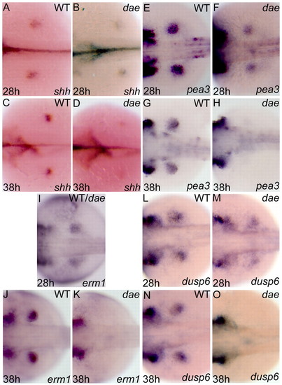
fgf10/dae mutant embryos have reduced expression of AER markers. Lateral views of wild-type (B,E,G,K,I,M,O,S,Q,U,W) and mutant fins (C,F,H,L,J,N,P,T,R,V,X), with anterior towards left. At 28 hpf, expression of fgf24 and bmp2b appears indistinguishable in wild-type and fgf10/dae (A,D). Expression of sp8 is weakly reduced in dae (H) but sp9 (L), dlx2a (P) and wnt3l (T) expression is strongly reduced in mutants. By 38 hpf, expression of all ridge markers analysed is reduced in fgf10/dae (C,F,J,N,R,V,X) when compared with siblings (B,E,I,M,Q,U,W). |
Expression of AER target genes is reduced in fgf10/dae. Dorsal views of in situ hybridisation staining of wild-type (A,C,E,G,J,L,N) and mutant embryos (B,D,F,H,K,M,O) with anterior towards the left. At 28 hpf, expression of all genes analysed appears either indistinguishable between fgf10/dae and sibling (I), or weakly reduced in mutant (B,F,M). By 38 hpf, expression of ridge target genes appear either strongly reduced (D) or absent (H,K,O) in fgf10/dae. |

Expression of DV axis markers is disrupted in fgf10/dae. Dorsal views of in situ hybridisation staining of wild-type (A,C,E,G) and fgf10/dae (B,D,F,H) embryos. Expression of all genes analysed appears slightly reduced at 28 hpf in fgf10/dae (B,F) when compared with siblings (A,E). By 38 hpf, eng1a expression is severely reduced (D) and wnt7a expression (H) is absent in fgf10/dae. |

Expression of fin marker genes compared in extl3/box and ext2/dak mutants. In situ hybridisation staining of wild-type (A,D,G,J), extl3/box (B,E,H,K) and ext2/dak (C,F,I,L) embryos. All panels are dorsal views with anterior towards the left. At 48 hpf, extl3/box mutant embryos have reduced expression of all markers analysed (B,E,H,K) when compared with wild type (A,D,G,J). ext2/dak mutants have a stronger phenotype. There is a strong reduction of dlx2a (C), fgf8 (F) and eng1a (L). Expression of shh is absent in ext2/dak (I) by 48 hpf of development. EXPRESSION / LABELING:
|
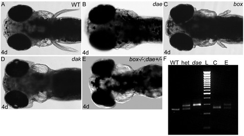
A strong genetic interaction between fgf10/dae and extl3/box during limb development. Four-day live photos (A-E) of wild-type (A), fgf10/dae (B), extl3/box (C), ext2/dak (D) and box–/–;dae+/– (E) embryos. All photos are dorsal views with anterior towards the left. dae (B) mutant embryos have a severe truncation of pectoral fin compared with siblings (A). extl3/box (C) mutants have a weaker pectoral fin truncation, whereas ext2/dak (D) appears similar to fgf10/dae and has a severe truncation of the fin. Removal of one copy of fgf10 in an extl3/box mutant background (E) severely worsens the extl3/box phenotype, demonstrating a genetic interaction between dae/fgf10 and box/extl3. SNP genotyping (F) confirms the dae/fgf10 genotype (see Materials and methods). WT, wild-type embryo; het, heterozygous embryo; dae, daedalus mutant embryo; L, ladder; C, box mutant embryo shown in C; E, box–/–;dae+/– embryo shown in E. |

Implantation of Fgf10 soaked beads into fgf10/dae mutant fin buds. Dorsal (A-E) and lateral (A′-E") views of 2.5-day-old embryos with anterior towards the left. Fgf10 protein rescues expression of fgf8 (A-A"), shh (B-B"), eng1a (C-C") and wnt7a (D-D") following bead implantation into the left-hand side fin bud (A′- D′) when compared with unoperated right-hand side fin buds (A"-D"). Yellow asterisks mark position of implanted beads (A′- E′). As a control, implantation of beads soaked in PBS (E-E") does not rescue marker gene expression. LV, left view of embryo; RV, right view of same embryo. |

Implantation of Fgf4 protein-soaked beads into fgf10/dae fin buds. Dorsal (A-C) and lateral (A′-C") views of 2.5-day-old embryos, anterior towards the left. Expression of genes indicated was examined by in situ hybridisation in operated left-hand side (A′- C′) and unoperated right hand side fin buds (A"- C"). Fgf4 protein is unable to rescue expression of fgf8 (A-A"). Conversely, Fgf4 protein is able to rescue the expression of both shh (B-B") and eng1a (C-C") in operated fin buds. Yellow asterisks indicate the position of implanted beads (A′- C ′). LV, left view of embryo; RV, right view of same embryo. |

Implantation of Fgf10-soaked beads into ext2/dak and extl3/box embryos. Dorsal (A-E) and lateral (A′-E") views of 2.5-day-old embryos, anterior towards the left. Expression of genes indicated was examined by in situ hybridisation in operated left-hand side (A′- E′) and unoperated right-hand side fin buds (A"-E") dak fin buds. Implantation of Fgf10 protein-soaked beads into extl3/box also fails to rescue the expression of shh (E′). Yellow asterisks indicate position of implanted beads (A′ - E′). LV, left view of embryo; RV, right view of same embryo. |

Implantation of Fgf4-soaked beads into ext2/dak and extl3/box embryos. Dorsal (A-C) and lateral (A′- C") views of 2.5-day-old embryos, anterior towards the left. Expression of genes was examined by in situ hybridisation in operated left-hand side (A′- C′) and unoperated right-hand side fin buds (A"-C"). Fgf4 protein beads strongly rescued the expression of eng1a (A-A") and wnt7a (B-B") in ext2/dak mutant embryos. Similarly, shh (C-C") expression was rescued in extl3/box following implantation of Fgf4-soaked beads. Yellow asterisks indicate position of implanted beads (A′- C′). LV, left view of embryo; RV, right view of same embryo. |