
- Title
-
Neuromuscular synapses can form in vivo by incorporation of initially aneural postsynaptic specializations
- Authors
- Flanagan-Steet, H., Fox, M.A., Meyer, D., and Sanes, J.R.
- Source
- Full text @ Development

Outgrowth of the CaP axon. (A-D) Cross-sections through a 20 hpf HB9-GFP fish counterstained with Neurotrace-435/455. (A) Position of the spinal cord (SC), motoneurons (MN), notochord (NC) and myotome (M). (B-D) Caudal (B), central (C) and rostral (D) segments illustrating extension of the CaP motor axon between the notochord and the ventral myotome. Multiple stages are seen in one fish because of the rostrocaudal developmental gradient. (E) Lateral view of six segments from an HB9:GFP fish at 24 hpf, showing the labeling of segmentally arranged primary motor axons. (F-K) Time-course of outgrowth in rostral segments. Motoneurons were present at 14 hpf (F). They began to extend processes tipped by filopodia (arrowheads) by 15 hpf (G), and formed stocky axons by 17 hpf (H). Initially large varicosities at 18 hpf (I) became interspersed with smaller varicosities by 22 hpf (J); varicosities become more uniform in size by 24 hpf (K). Axons were labeled by GFP in HB9:GFP fish (A-H,J) or by the znp-1 antibody in wild-type fish (I,K). (L) Schematic of CaP axon outgrowth (not to scale). Lavender band indicates the position of cells that are, or will become, muscle pioneers. Scale bar in F: 10 µm for F-K. |

AChR clustering precedes axon outgrowth. CaP axons (green) labeled with znp-1 (B,C) or GFP (A,D-I) as in Fig. 1; AChRs labeled with BTX (red). (A-D) Segments 1-3. Neither axons nor AChR clusters are present at 14 hpf (A), but clusters are present in front of the advancing axon by 16-17 hpf (B,C). By 20 hpf, (D) most AChR clusters are apposed by axonal varicosities, but some clusters are larger than varicosities (arrowheads) and some axonal processes extend beyond clusters (arrow). Asterisks in B-D indicate some of the AChR clusters that are not innervated; they are quantified in Fig. 6F. (E-I) Whole mount of a 20 hpf fish (E), and higher-power details of individual segments (F-I) showing the rostrocaudal developmental sequence (see also Fig. 1A). In caudal segments (F), AChR clusters have formed but no axon has yet extended. In intermediate segments (G,H), clusters on adaxial cells not contacted by CaP disperse (asterisks in E). In the most rostral segments (I), the CaP axon has extended beyond the ventral-most AChR clusters. Scale bars: in A, 10 µm for A-D; in E, 25 µm; in I, 10 µm for F-I. |
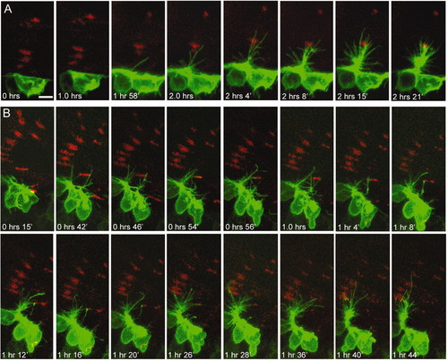
CaP axons incorporate initially aneural AChR clusters into NMJs. (A,B) HB9:mGFP fish were counterstained with BTX (red) and imaged every 2 minutes for 2 hours starting at 17 hpf (0 hours). In both A and B, AChR clusters were present before filopodia approached them. The axon in B initially pioneered an incorrect path toward a group of clusters caudal to the end-plate band. Later, it retracted and then took a path down the central band. Dorsal is down and rostral to left in all panels. Scale bar: 10 µm. |
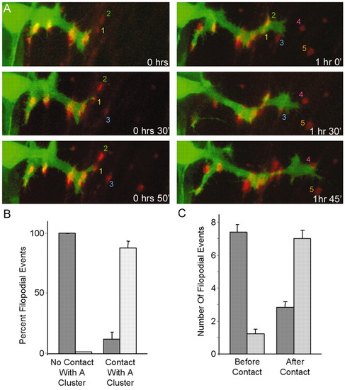
CaP axonal behavior changes following contact with AChR clusters. (A) HB9:GFP-labeled CaP reorients its growth repeatedly following contact of its growth cone with clusters. Colored numbers 1-5 indicate initially aneural clusters that growth cones contact. (B) Most of the filopodia that contact an AChR cluster are retained (light bars) until the end of the imaging period, whereas most of those that do not make contact retract (dark bars). (n=173 filopodia from 13 cells in seven animals; P<0.0001 for both groups.) (C) Filopodial activity is lower in the quadrant surrounding the central band of AChRs (light bars) than in the sum of the other three quadrants (dark bars), prior to the initial contact of a filopodium with a cluster. After the first filopodium contacts a cluster, however, more filopodia extend in the quadrant containing the end-plate band than in the other three quadrants together (n=1022 filopodia from 10 cells in five fish; P<0.0001 both before and after contact). |
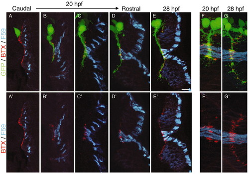
Distinct modes of synapse formation on muscle pioneers and fast fibers. Cross-sections through HB9:GFP fish at 20 hpf (A-D,F) or 28 hpf (E,G) were stained with anti-myosin (F59) and BTX. (A-D) CaP extends to contact muscle pioneers as the other adaxial fibers migrate radially. (E) CaP then extends past the pioneers as fast, weakly F59-immunoreactive, fibers form. AChR clusters on migrating adaxial cells are lost and new clusters form on fast fibers as, or after, they are contacted by CaP. As in Figs 1 and 2, the rostrocaudal sequence of development allows the visualization of many stages in a single fish. (F,G) En face views at 20 hpf and 28 hpf to show that AChR clusters on muscle pioneers are larger and more elongated than those on fast fibers. A'-G' show F59 and BTX, but not axons, to highlight AChR cluster position and morphology. Scale bar in E: 10 µm for all panels. |

Apposition of CaP axon varicosities to AChR clusters. (A-C) Axons and AChRs labeled with znp-1 (green) and BTX (red), respectively. By 20 hpf, axonal varicosities are apposed to AChR clusters, but some clusters are larger than varicosities (arrowhead) and some axonal processes extend beyond clusters (asterisk). Yellow asterisks indicate AChR clusters that are not innervated. Apposition becomes precise by 24 hpf (B,C). At this time, pre- and postsynaptic specializations are perfectly aligned (C), varicosities are centered on myotubes (D; phalloidin in blue) and each myotube bears a single AChR cluster (E; single myotube labeled with mYFP by mosaic expression of a transgene injected into a one-cell embryo). The inset in E is a higher-power view of the single cluster indicated by the asterisk. (F) Progressive loss of aneural clusters. Aneural clusters lying rostral or caudal to CaP disperse (solid line), whereas many of the aneural clusters in front of the axon (dashed line) are incorporated into NMJs (n=10-64, average=38, segments per stage). (G) Cluster size decreases between 20 and 24 hpf (P<0.0001 by t-test; n=250 clusters per stage). Scale bar in C: 5 µm for A-E. |

AChR clusters form but do not mature or persist in the absence of axons. (A) An HB9:mGFP+/+ fish at 24 hpf. Out of six segments shown (asterisks), axons extend in only two. (B-E) HB9:mGFP+/+ fish at 19 (B), 22 (C), 24 (D) and 48 (E) hfp, counterstained with BTX (red). Bands of AChR clusters formed in segments that were not innervated, but did not persist. AChRs did cluster following outgrowth of secondary axons (E). Asterisks in B indicate aneural clusters; asterisk in D indicates muscle pioneers without clusters. (F) Analysis of cluster number in segments that were not innervated (Un) and in innervated segments (In) of HB9:mGFP+/+ fish. Segments that were not innervated contained the same number of clusters as innervated segments at 19 hpf, but contained nearly twice as many by 22 hpf. In the absence of innervation, most clusters disperse by 24 hpf (n=10-35 segments per bar, average=16; P=0.82 at 19 hpf, P<0.0001 at 22 hpf, P<0.0001 at 24 hpf). (G) Clusters present in innervated segments and in segments that were not innervated were similar in length at 19 and 22 hpf, but differed at 24 hpf (n=30-102 clusters per bar, average=60; P=0.31 at 19 hpf, P=0.92 at 22 hpf, and P<0.05 at 24 hpf). Scale bar in B: 10 µm for A-E. |