
- Title
-
Molecular characterization and embryonic expression of the family of N-methyl-D-aspartate receptor subunit genes in the zebrafish
- Authors
- Cox, J.A., Kucenas, S., and Voigt, M.M.
- Source
- Full text @ Dev. Dyn.

A: Polymerase chain reaction (PCR) reaction using cDNA from 4-day-old zebrafish. The forward primer lies in exon 4, and the reverse primer in exon 6 of zNMDA R1.1 and zNMDA R1.2 The larger band represents a splice variant, which contains the exon 5 insertion, and the smaller band the splice variant, which lacks this insertion. B: PCR reaction as in A. This strategy represents the C-terminal splice variants produced using a forward primer in exon 20 and a reverse primer in exon 22 of NR1.1 and NR1.2. The larger band represents the splice variant containing exon 21 and the smaller band lacks this exon. C: A diagram representing the three splice variants of zNR1.1, which we detected in zebrafish. ZNR1.2 displays the same splice variants. EXPRESSION / LABELING:
|
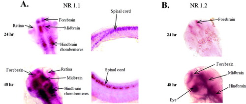
A: Gene expression of NR1.1 in the head and spinal cord of 24 hours postfertilization (hpf) and 48 hpf. At 24 hpf, NR1.1 is highly expressed in the fore-, mid-, and hindbrain; in the retina; and in the spinal cord. The 24 hpf spinal cord is slightly flexed, presenting a more dorsal view at the rostral end (left), showing bilateral cell labeling. More caudally, the specimen shows that the positive neurons are found both ventrally (top) and dorsally (bottom) in the cord. At 48 hpf, this expression pattern increases in intensity in these same tissues. B: Gene expression of NR1.2 paralog. At 24 hpf, there is light staining in the forebrain. By 48 hpf, all three regions of the brain express NR1.2. The 48 hpf image is a dorsolateral view of the left side of the head. This paralog is not expressed in the spinal cord. |
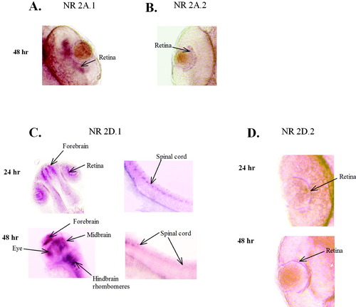
A: Gene expression of NR2A.1 in the retina of 48 hpf zebrafish. This subunit is not expressed at 24 hours postfertilization (hpf). B:Expression of NR2A.2 in the retina of 48 hpf but not 24 hpf zebrafish. C:In situ hybridization pattern of the NR2D.1 RNA in 24- and 48-hour zebrafish. At 24 hpf, this subunit is found in the forebrain, retina, and spinal cord. The orientation of the spinal cord is rostral to the left, caudal to the right, dorsal at the top, and ventral at the bottom. At 48 hpf, the expression has increased in intensity and is found in the fore-, mid-, and hindbrain, and in the spinal cord. The photo of the head at 48 hpf is a dorsolateral view. This expression pattern overlaps that of NR1.1. D: NR2D.2 is detectable in the retina at 24 and 48 hpf but is not found in the brain or spinal cord. All images were cropped and sized in Photoshop 6.0. |

Polymerase chain reaction (PCR) reaction using cDNA from 24 hpf, 48 hpf, 96 hpf, and 2-month-old zebrafish embryos. For zN2B.1, the forward primer lies in exon 12 and the reverse primer lies in exon 15, yielding a fragment of 680 bp. For zN2B.2, the forward primer lies in exon 4 and the reverse primer in exon 6, yielding a fragment of 775 bp. The N2B.1 does not appear to be expressed at 24 or 48 hpf but is seen at 96 hpf. The N2B.2 is not expressed at 24 hpf. It appears to be expressed at very low levels at 48 hpf and is strongly expressed at 96 hr. The expression has decreased by 2 months after fertilization. EXPRESSION / LABELING:
|