
- Title
-
Hedgehog signaling is required for cranial neural crest morphogenesis and chondrogenesis at the midline in the zebrafish skull
- Authors
- Wada, N., Javidan, Y., Nelson, S., Carney, T.J., Kelsh, R.N., Schilling, T.F.
- Source
- Full text @ Development
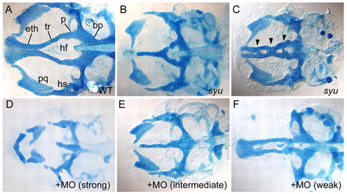
Neurocranial cartilage patterns in wild-type and Hh-deficient larvae. Alcian stained cartilages were dissected and flat mounted; dorsal views are shown, anterior to the left. (A) The wild-type ANC at 4.5 dpf includes paired trabeculae (tr) and an ethmoid plate (eth). Mandibular (pq) and hyoid (hs) cartilages remain attached. (B,C) Neurocranial defects in syu mutants. Trabecular cartilages either fuse completely (B) or partially (C, arrowheads) at the midline. (D-F) Concentration-dependent effects of co-injected shh-MO and twhh-MO in wild type: (D) 3.0 ng shh-MO + 0.82 ng twhh-MO; (E) 2.0 ng shh-MO + 0.55 ng twhh-MO; (F) 1.0 ng shh-MO + 0.27 ng twhh-MO. bp, basal plate; eth, ethmoid plate; hf, hypophysial fenestra; hs, hyosymplectic; pq, palatoquadrate; p, polar cartilage; tr, trabeculae. |

Early chondrogenesis in the ANC. (A-H) Alcian Blue-stained embryos. (I-P) In situ hybridization for sox9a mRNA. Ventral views in wild type (A-C) and in syu mutants (E-G) between 48-56 hpf are shown. (A) Paired trabeculae consist of single rows of chondrocytes. (B,C) These elongate (B) and fuse posteriorly (C) to the parachordals. (D) Lateral view, 56 hpf, showing the ventral position of trabeculae (dotted line). (E) In syu mutants, the first ANC cartilages (dotted lines) are shorter and closer to the notochord (arrowhead). (F) Trabeculae fuse at the midline and (G) this fusion persists. (H) A lateral view of a syu mutant at 56 hpf. (I-K) Ventral views of sox9a expression in wild type between 30-45 hpf. (M-O) sox9a expression in syu mutants. (I) Early expression in bilateral cell clusters (arrowheads) adjacent to the diencephalon (Di). (J,K) These elongate posteriorly (J), and fuse in the midline anteriorly (K). (L) Lateral view of wild type at 45 hpf. (M) In syu mutants, sox9a expression is delayed. (N,O) sox9a+ clusters elongate (N) and fuse anteriorly (O, arrow). (P) Lateral view of sox9a expression in syu mutants showing fused trabeculae (dotted lines). Asterisk indicates anterior extent of expression. eth, ethmoid plate; MC, Meckel's cartilage; tr, trabeculae. Scale bar: 50 μm. |

Expression of the sox10:egfp transgene in cranial NC cells of living embryos. (A-C) Representative images from a confocal, time-lapsed movie of sox10:egfp expression in the head between 14-20 hpf, lateral view (see also Movie 1 in the supplementary material). (A-C) Brightfield (A) and confocal images (B,C) at 14 hpf (A,B) and 20 hpf (C). (D) Diagram illustrating migration paths of individual NC cells, traced by analyzing confocal stacks within the movie. Circles indicate premigratory cell positions and stars indicate their positions six hours later. Some cells (dark green triangle) could not be tracked beyond 17.5 hpf, whereas others (light green square) were only traceable after 16.5 hpf. (E-N) sox10:egfp+ cells at later stages in wild-type (E-I) and Hh-deficient (J-N) embryos. (E) Lateral view at 24 hpf, showing the first pharyngeal arch (PA1) and stomodeum (arrowhead). Precursors of the ANC lie beneath the eye and anterior to this stomodeal pouch. (F,G) Ventral views with (G) and without (F) accompanying brightfield images. (H,I) By 36 hpf, sox10:egfp+ cells aggregate around the mouth (H), and, by 48 hpf, form cartilage of the ANC (I). (J) In Hh-deficient embryos, sox10:egfp expression is similar to that seen in wild-type controls, when viewed laterally. (K,L) However, in ventral view the head is mediolaterally compressed, and NC cells anterior to PA1 fuse across the ventral midline, between the eyes (arrow). (M) Fusion is more pronounced at 36 hpf, forming a single midline condensation (arrow). (N) A single rod of sox10:egfp+ cartilage forms in the midline by 48 hpf. Scale bar: 50 μm. |

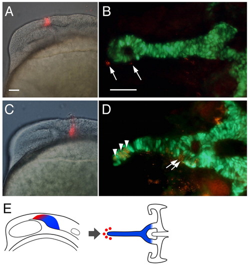
Fate maps of NC contributions to the ANC. (A,C,E,G) Labeled cells immediately after injection of PKH26, seen in lateral views of living embryos at 13 hpf. (B,D,F,H) Labeled cartilage in the ANC at 80-85 hpf, showing colocalization (yellow) of PKH (red) and sox10:egfp (green), ventral view. (A) Premigratory cranial NC dorsal to the optic vesicle forms median ethmoid (B, arrows). (E) Premigratory NC posterior to the optic vesicle forms trabeculae (F, arrowheads). (C) Postmigratory NC cells anterior to the optic stalk at 22-24 hpf form median ethmoid (D, arrows). (G) Postmigratory NC posterior to the optic stalk form trabeculae (H, arrowheads). (I,J) Expression of sox10 (I) and foxd3 (J) mRNA at 13 hpf, lateral view. Lines delineate the posterior edge of the optic vesicle. (K) Schematic representation of the fate map. In premigratory NC, ethmoid precursors (red) migrate anteriorly, trabecular precursors (blue) migrate laterally and ventrally. Scale bars: 50 μm; in A for A,C,E,G,I,J; in B for B,D,F,H. EXPRESSION / LABELING:
|
Fate maps in Hh-deficient embryos. (A,C) PKH labeling immediately after injection, lateral views. (B, D) Labeled cells at 80 hpf derived from these injections, showing co-localization (yellow) of PKH (red) and sox10:egfp (green) in dissected preparations, ventral views. (A) NC cells labeled dorsal to the optic vesicle do not contribute to cartilage, but remain undifferentiated (B, arrows). (C) By contrast, cells labeled posterior to the optic vesicle contribute to the midline cartilage rod (D, arrowheads) and posterior trabeculae (arrows). (E) Schematic representation of the fate map in Hh-deficient embryos. Trabecular precursors form cartilage (blue), whereas ethmoid progenitors (red) do not. Scale bars: 50μm; in A for A,C; in B for B,D. |
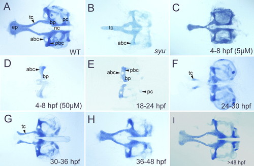
CyA treatment disrupts ANC formation. Flat-mounted, Alcian Blue stained cartilages at 5 dpf; anterior is to the left. (A) Wild type. (B) syu mutant. (C) A wild-type treated with 5 μM CyA from 4-8 hpf. (D-I) Wild-type larvae treated with 50 μM CyA at different stages between 18-60 hpf. (D,E) Treatment between 4-24 hpf eliminates all ANC cartilage. (F) Treatment between 24-30 hpf eliminates most cartilage, although some trabecular cartilage forms but never fuses to the posterior neurocranium. (G) Treatments between 30-36 hpf eliminate the ethmoid, but not trabeculae. (H,I) Treatments later than 40 hpf cause slight reductions in the ANC. abc, anterior basicapsular commissure; bp, basal plate; ep, ethmoid plate; nc, notochord; pbc, posterior basicapsular commissure; pc, parachordal cartilage; tc, trabeculae. |
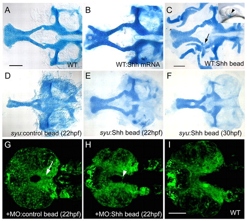
Exogenous Shh expands ANC cartilage in wild types and rescues syu mutants. Ventral views, anterior to the left. (A-F) Flat-mounted, Alcian-Blue-stained cartilages at 4.5 dpf. (G-I) Confocal images of sox10:egfp expression in dissected embryos at 36 hpf. (A) Wild type. (B) Wild type injected with shh mRNA. Trabeculae expand laterally and often separate from the ethmoid (asterisk). (C) Wild type implanted with a Shh-coated bead on the left side at 20 hpf. The ANC expands near the bead; both trabeculae and ethmoid thicken, and the trabecula on the implanted side fuses to the palatoquadrate (arrow). Inset shows the typical bead position (arrowhead). (D) Control beads implanted at 22 hpf had no effect on ANC formation in syu mutants. (E) Implantation of a Shh-coated bead partially rescues the ANC in syu mutants. (F) Bead implantation at 30-36 hpf also partially rescues the ANC in some cases. (G) Control beads implanted between 22-36 hpf had no effect on the distribution of sox10:egfp+ cells in Hh-deficient embryos. Arrow indicates NC cells at the midline. (H) Implantation of a Shh-coated bead at 22 hpf rescued sox10:egfp expression at the ventral midline (arrowhead). Note that the width of the bead-implanted embryos is identical to that of controls (G,H), and smaller than that of wild type (I). Scale bars: 50 μm; in A for A,B; in C for C-F; in I for G-I. |

Shh is required in both ventral neural tube and oral ectoderm for ANC formation. (A-C,E,F,H,I) Flat-mounted Alcian-Blue stained cartilage at 5 dpf; anterior is to the left. (D,G) Lateral views of transplants shown in E and H, prior to cartilage dissection. Transplanted donor cells (brown) lie in the ventral brain. (A) Wild type. (B) Partial rescue of the ANC by wild-type ectoderm transplanted into a syu mutant host. Transplanted cells lie close to the ethmoid plate in this example. (C) syu control sibling of the embryo shown in B. (D) Grafted wild-type cells in the ventral forebrain of a syu mutant. (E) Partial rescue of the ANC by the transplant shown in D. Grafted cells were lost during dissection. (F) syu control sibling of embryo shown in E. (G) An shh-MO/twhh-MO-injected embryo. Inset shows transplanted cells at 28 hpf. (H) Partial rescue of ANC in the embryo shown in G. (I) shh-MO/twhh-MO-injected control sibling of embryo in H. abc, anterior basicapsular commissure; ep, ethmoid plate; nc, notochord; pbc, posterior basicapsular commissure; pc, parachordal cartilage; tc, trabeculae. |