
- Title
-
A mutation in the silver gene leads to defects in melanosome biogenesis and alterations in the visual system in the zebrafish mutant fading vision
- Authors
- Schonthaler, H.B., Lampert, J.M., von Lintig, J., Schwarz, H., Geisler, R., and Neuhauss, S.C.
- Source
- Full text @ Dev. Biol.

Body pigmentation of the fading vision mutant. (A) 5-day-old wild-type and (B) fdv mutant larva. fdv shows lighter body pigmentation compared to the 5-dpf-old wild-type larva. (C, E) Higher magnification reveals that the shape of the pigment cells and the distribution of melanin in the pigment cells are different between (C) 5-dpf-old wild-type larvae and (E) 5-dpf-old fdv mutant larvae. (Arrowhead in panels C and E) Some pigment cells appear almost unpigmented. Hypopigmentation is clearly apparent in panel (D) fins of adult wild-type fish compared to (F) homozygous adult fdv mutants. Scale bars in panels (A) and (B) are 500 μm. Scale bars in panels (C) and (E) are 200 μm. Scale bars in panels (D) and (F) are 500 μm. |

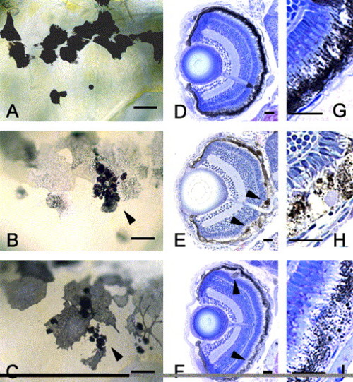
Histological eye sections at different developmental stages. Comparisons of (A–E) wild-type sections with (F–J) mutant sections show a progressive morphological deterioration in the outer layer of the retina. At 3 dpf, the RPE appears to be lighter pigmented in (F) fdv mutants than in (A) wild-type larvae. (B and G) Sections taken from 5-dpf-old larvae show a thickening of the RPE in the mutant larvae, (arrowheads in G) the appearance of pigment clots in the RPE and a reduced length of the outer segments in fdv mutants, while the (B) wild-type RPE is densely pigmented and microvilli of the RPE interdigitate with the PRC outer segments. In sections of (H) 7 dpf fdv larvae, the RPE is even more severely affected. The RPE cells are lighter pigmented in the mutant, containing pigment clots and no microvilli. PRC outer segments are bent sideways, as can be seen at higher magnification ((D) wild-type and (I) fdv mutant) and (arrowhead in panel I) are even absent in some parts of the mutant retina. Surprisingly, a comparison of sections of (J) adult fdv mutant and (E) wild-type eyes shows that the RPE and the PRC outer segments have recovered in the mutant, and (arrowheads in panels E and J) microvilli interdigitating in between the outer segments of PRCs can be seen. The inner retina is not affected at any of the developmental stages examined. ON, optic nerve, GCL, ganglion cell layer, IPL, inner plexiform layer, INL, inner nuclear layer, OPL, outer plexiform layer, ONL, outer nuclear layer, OS, outer segments, RPE, retinal pigment epithelium. Scale bars in panels (A–C), (F–H), (E) and (J) are 50 mm. Scale bars in panels (D) and (I) are 20 mm. |

Transmission electron microscopy from wild-type and fdv. Ultrathin sections were taken from (A, E) 3 dpf, (B, C, F, G) 5 dpf and (D, H) adult zebrafish. At larval stages (A–G), PRC outer segments in fdv mutants are significantly shorter and the RPE is damaged. In the RPE of fdv mutants, vacuoles begin to form at (E) 3 dpf and are prominent at (asterisks in panel F) 5 dpf. Melanin-containing pigment granules are aberrantly shaped. (C and G) Organelles containing detached PRC outer segments can be observed more often within RPE cells of fdv mutants than in wild-type larvae. Panel (G) shows a higher magnification of the box depicted in panel (C). The arrows in panels (C) and (G) indicate the undigested stacks of PRC outer segments in the RPE. The microvilli that interdigitate between the PRC outer segments (arrow in panel B) in wild-type larvae are rarely found in larvae of fdv mutants. (D, H) In adult fdv, the outer segments have recovered, but the melanosomes contain vesicles and some have a fuzzy diffuse shape. OS, outer segments of photoreceptor cells. The thin black lines in panels (B), (E) and (F) are due to preparation artifacts. Scale bars in panels (A) and (E) are 2 μm, in panels (B) and (F) 10 μm, in panel (C) 1 μm, in panel (G) 0,5 μm, in panels (D) and (H) 2 μm. |

Spatio-temporal expression pattern of silva and silvb. Expression analyses in 2-day-old wild-type larva. (A and B) silva is expressed in (arrowheads in A) melanocytes and (B) the RPE of the eye. (C) On adult sections, the silva transcript is confined to cells of the RPE and to the melanocytes located behind the eye. (D and E) silvb expression is restricted to RPE cells. RPE, retinal pigment epithelium, PRC, photoreceptor outer segments, ONL, outer nuclear layer, OPL, outer plexiform layer, INL, inner nuclear layer, IPL, inner plexiform layer, GCL, ganglion cell layer. Scale bar in panel A is 50 µm, in panels B and D 100 μm, in panel C 20 μm and in panel E 50 μm. |
Phenocopy of fdv by silva knock-down. (C, F, I) Morpholino-oligonucleotide-injected 5-day-old larvae show the same phenotype as (B, E, H) 5-day-old fdv mutant larvae. Compared to the (A, D, G) wild-type, the melanocytes are lighter and contain pigment clots (arrowheads in panels C and F). In the sections taken from (arrowheads in panel F) larvae after morpholino injection, some RPE cells are bloated and contain vacuoles as can be seen in (arrowheads in panel E) fdv mutant larvae. Scale bars in panels A–C are 20 μm, in panels D–F 100 μm and in panels G–I 50 μm. |
Reprinted from Developmental Biology, 284(2), Schonthaler, H.B., Lampert, J.M., von Lintig, J., Schwarz, H., Geisler, R., and Neuhauss, S.C., A mutation in the silver gene leads to defects in melanosome biogenesis and alterations in the visual system in the zebrafish mutant fading vision, 421-436, Copyright (2005) with permission from Elsevier. Full text @ Dev. Biol.