
- Title
-
Cooperative function of deltaC and her7 in anterior segment formation
- Authors
- Oates, A.C., Mueller, C., and Ho, R.K.
- Source
- Full text @ Dev. Biol.
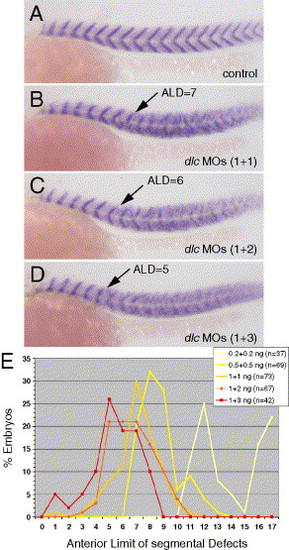
Effect of reduction of deltaC function on zebrafish segmentation. (A–D) Myotome boundaries of the trunk marked by titin expression are shown in 26 hpf embryos in lateral view with anterior to the left and dorsal upwards. Arrows in panels (B–D) indicate Anterior Limit of Defects (ALD) in each embryo. (A) Uninjected control. Representative embryos injected with 1 ng/nL (B), with 1 and 2 ng/nL (C) and with 1 and 3 ng/nL (D) of each of the morpholinos dlcMO3 and dlcMO4, respectively. (E) Data from a histogram (plotted as line graph) showing the distribution of ALD for populations of embryos injected with increasing concentrations of combined morpholinos, indicating a final ALD at segment 5. Embryos injected with doses higher than 1 + 3 ng/nL of the combined morpholinos exhibited necrosis and non-specific defects and were not included. EXPRESSION / LABELING:
|

Effect of reduction of deltaC function on somitogenesis and segment polarity. Gene expression patterns in PSM and trunk somites of embryos at 14 hpf (10 somites) are shown in dorsal view after flat mounting with anterior upwards. In each pair of panels (A–D, F, H–J), an uninjected control is on the left and an embryo coinjected with 1 ng/nL dlcMO3 and 2 ng/nL dlcMO4 is on the right. In panels (E) and (G), the uninjected embryo is on the left (a) and embryos injected with dlcMOs (b), her7MOs (c; 2 ng/nL her7MO1 and 2 ng/nL her7MO2), or homozygous for the aei/dld mutation (d) are ordered to the right. Expression of myoD (A), fgf8 (B), fgfr4 (C), notch6 (D), notch5 (E), mespb (F), mespa (G), papc (H), ephA4 (I), and ephrinB2 (J) is shown. Brackets in panels (Ed) and (I) and arrow in panel (Ec) indicate affected regions, asterisks in panel (E) mark expression in overlying neural plate. EXPRESSION / LABELING:
|
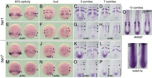
Expression of the cyclic her genes in response to reduction of deltaC function. Expression in PSM of zebrafish cyclic genes her1 (A–H) and her7 (I–P) shown from 9 hpf (80% epiboly) to 13 hpf (7 somites). Expression of non-cyclic deltaD (dld; Q, R) and notch1a (S, T) shown at 14 hpf (10 somites). Embryos are displayed in whole mount (A, B, E, F, I, J, M, N) and in flat mount (C, D, G, H, K, L, O, P, Q–T) with (E–H, M–P, R, T) and without (A–D, I–L, Q, S) injection of 1 ng/nL dlcMO3 and dlcMO4. In each panel (A–D, I–L), two wild type embryos with representative phases of cyclic gene expression patterns are shown; wavelike expression domains are marked with asterisks. Batches of dlcMO-injected embryos show similar variability to wild type embryos at earlier stages (E, M), but at later stages, embryos in a batch are indistinguishable (H, P). Regions of invariant gene expression are denoted with brackets in panels (O), (H), and (P) and asterisks in panel (P). Note also that, across this time period, the level of transcript decreases in dlcMO-injected embryos. (Q, R) The effect of dlcMOs (as above) on the expression of deltaD showing a loss of patterning in the presence of normal levels of dld expression. (S, T) Expression levels of notch1a are not perturbed by injection of dlcMOs. EXPRESSION / LABELING:
|
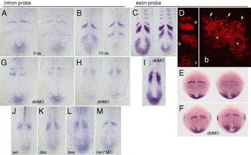
Transcriptional activity of the deltaC gene in wild type and segmentation defective mutant backgrounds. Distribution of total dlc mRNA, detected with a cDNA-derived riboprobe, is shown in panels (C–F) and (I). Distribution of unspliced dlc pre-mRNA, detected with a riboprobe to the fourth intron, is shown in panels (A), (B), (G), (H), and (J–M). Dorsal view in flat mounted embryos in wild type (A–E), morpholino injected (F–I.M), and mutant (J, aei/dld; K des/n1a,; L, bea) backgrounds. Embryos are shown at bud (E, F), 3 (A, G), 7 (D), and 10 somite (B, C, H, I, J–M) stages of development. Panels (F–I) show the effect of injection of 1 ng/nL dlcMO3 and 2 ng/nL dlcMO4, and panel (M) shows the effect of 2 ng/nL her7MO1 and her7MO2. The total distribution of dlc mRNA in a representative embryo, as detected with the exonic riboprobe by confocal LSM, is shown in panel D, with (a) anterior, (b) mid-, and (c) posterior wavelike expression domains, (b) and is magnified in right panel showing puncta (arrows) on the rostral side, and diffuse staining (asterisks) in the caudal part of the expression domain. EXPRESSION / LABELING:
|
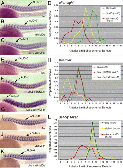
Analysis of the interaction between deltaC and the after eight/deltaD (aei/dld), beamter (bea), and deadly seven/notch1a (des/n1a) mutations in the Delta/Notch signaling pathway. Myotome boundaries in the trunk are shown in 26 hpf embryos in lateral view with anterior to the left and dorsal upwards (A–C, E–G, I–K). (A) aei/dld uninjected, (B) wild type injected with 1 ng/nL dlcMO3 and 1 ng/nL dlcMO4, (C) aei/dld injected with dlcMOs. (E) bea uninjected, (F) bea injected with 1 ng/nL dlcMO3 and 2 ng/nL dlcMO4, (G) bea injected with 3 ng/nL her7MO1 and 3 ng/nL her7MO2. (I) des/n1a uninjected, (J) wild type injected with 0.5 ng/nL dlcMO3 and 0.5 ng/nL dlcMO4. (D, H, L) Histograms comparing Anterior Limit of Defects (ALD) for these nine conditions. Arrows in panel (A–C), (E–G), and (I–K) indicate ALD in each embryo. EXPRESSION / LABELING:
|

Analysis of the interaction between deltaC and her7. Myotome boundaries in the trunk are shown in 26 hpf embryos in lateral view with anterior to the left and dorsal upwards (A, B). (A) Wild type, uninjected control, (B) injected with 3 ng/nL her7MO1 and 3 ng/nL her7MO2. (C) Histogram comparing ALD in populations of embryos injected with increasing amounts of combined her7MOs, where the two numbers (e.g. 1 + 1) in inset legend are the concentration of her7MO1 and her7MO2 morpholino, respectively in ng/nL. Embryos injected with doses higher than 3 + 3 of combined morpholinos exhibited necrosis and non-specific defects. (D) Wild type embryo injected with 2 ng/nL her7MO1 and 2 ng/nL her7MO2, (E) embryo injected with 1 ng/nL dlcMO3 and 2 ng/nL dlcMO4, (F) embryo injected with her7- and dlcMOs combined. Arrows in panels (A), (B), and (D–F) indicate Anterior Limit of Defects (ALD) in these embryos. (G) Histogram comparing ALD in populations of embryos injected with the treatments in panels (D–F). (H–O) Expression of the her1 cyclic gene after injection of dlc- and her7MOs. Presomitic mesoderm of embryos at 80% epiboly (H, J) and bud (I, K) in whole mount. Comparison of coherent wavelike expression domains of her1 expression in wild type (H, I) and her7- and dlcMO-injected embryos (J, K). In each panel (H, I), two wild type embryos with representative phases of cyclic gene expression patterns are shown. In contrast, her7- and dlcMO-injected embryos are indistinguishable from each other (J, K). (L–O) Expression of the cyclic gene her1 in the PSM of embryos at 14 hpf (10 somites) in dorsal view after flat mounting with anterior up. (L) Two representative phases of cyclic her1 expression in wild type, control embryo; (M) comparison of levels and patterns of her1 expression after injection of dlcMOs (1 ng/nL dlcMO3 and 2 ng/nL dlcMO4), (N) dlc- and her7MOs at the concentrations mentioned, (O) her7MOs (2 ng/nL her7MO1 and 2 ng/nL her7MO2). For the treatments shown in panels (M–O), embryos in a batch were indistinguishable from each other. EXPRESSION / LABELING:
|
Reprinted from Developmental Biology, 280(1), Oates, A.C., Mueller, C., and Ho, R.K., Cooperative function of deltaC and her7 in anterior segment formation, 133-149, Copyright (2005) with permission from Elsevier. Full text @ Dev. Biol.