
- Title
-
A homologue of the Drosophila kinesin-like protein Costal2 regulates Hedgehog signal transduction in the vertebrate embryo
- Authors
- Tay, S.Y., Ingham, P.W., and Roy, S.
- Source
- Full text @ Development

Expression pattern of the zebrafish cos2 gene and subcellular distribution of the Cos2 protein. (A) cos2 expression in a 12-somite stage embryo derived from a cross of heterozygous smub641 parent fish. The expression is indistinguishable in wild-type embryos and their homozygous mutant smu siblings. (B) cos2 expression in a 12-somite stage wild-type embryo injected with Shh mRNA. (C) Flat mount of a 15 somite wild-type embryo with uniform expression of cos2 in somitic cells (arrows) and the midline (asterisk). (D) Lateral view of the myotome of a wild-type embryo at 24 hours post fertilisation (hpf) showing cos2 expression in muscle fibres. (E) Somitic cells of a two- to three-somite stage wild-type embryo injected with GFP-Cos2 RNA exhibiting cytoplasmic distribution of the fusion protein (n=5/5). (F) Superimposition of the GFP channel depicted in E with that of ToPro-3 fluorescence to highlight the nuclei. (G) Somitic cells of a two- to three-somite stage wild-type embryo injected with GFP-Cos2 RNA and stained with antibodies to ß-tubulin showing the cytoplasmic distribution of microtubules. (H) Superimposition of the image in G with the pattern of GFP-Cos2 distribution reveals substantial overlap of the GFP signal and the microtubules. Panels illustrating embryos in this and subsequent figures are oriented anterior towards the left and dorsal towards the top, unless mentioned otherwise. EXPRESSION / LABELING:
|
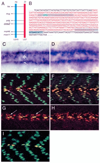
Loss of Cos2 function results in de-repression of the Hh pathway in the myotome of the zebrafish embryo. (A) Diagrammatic representation of the synteny between regions of human (Hs; LG15) and zebrafish (Dr; LG7) chromosomes bearing the cos2 genes. fes, proto-oncogene tyrosine protein kinase; rhcg, Rh type-C glycoprotein; polg, DNA polymerase subunit 1; mrpl46, mitochondrial ribosomal protein L46; mrps11, mitochondrial ribosomal protein S11. (B) Nucleotide sequence of the first protein coding exon of cos2 and the flanking introns. The start codon is indicated in green. The sequences targeted by the start and splice MOs are highlighted. (C) Flat mount of a two-somite stage wild-type embryo, showing the pattern of ptc1 transcription. The precursors of the SSFs and MPs (arrows) and the neural plate (asterisk) are indicated. (D) Similar flat mount of a cos2 morphant with expanded domain of ptc1 expression in the somitic mesoderm (n=9/16). (E) Lateral view of the myotome of a 24 hpf wild-type embryo showing the SSFs (stained for expression of the homeodomain protein Prox1, green) and the MPs [stained for expression of the homeodomain proteins of the Engrailed (Eng) family, red]. Within the slow muscle lineage, Prox1 is expressed in SSFs as well as MPs, while Eng proteins are expressed exclusively in the MPs. Prox1- and Eng-positive MP nuclei (yellow) are indicated (arrows). (F) A similar stage cos2 morphant with supernumerary MP cells in the myotome (n=6/7). (G) Medial view of the myotome of a 24 hpf wild-type embryo. MFFs that surround the MPs and express low levels of Eng are indicated (arrows). (H) MFFs are increased in numbers in cos2 morphants (n=5/7). (I) Myotome of an embryo co-injected with cos2 MOs and cos2 sense mRNA, showing effective suppression of supernumerary MP induction that is observed in cos2 morphants (n=12/12). EXPRESSION / LABELING:
PHENOTYPE:
|

Inactivation of Cos2 function induces ectopic Hh signalling in the ventral neural tube. (A) fkd4 expression in the ventral neural tube (MFP and LFP cells) of a 22-somite stage wild-type embryo. (B) Upregulation of the levels and expansion in the domain of fkd4 expression in the ventral neural tube of a 22-somite stage cos2 morphant (n=16/22). (C) Expression pattern of fkd4 in ventral cell populations of the developing mid- and hindbrain of a 22-somite stage wild-type embryo. (D) Increased levels and enlargement of the domain of fkd4 in ventral cell populations of the developing mid- and hindbrain of a 22-somite stage cos2 morphant embryo (n=13/22). EXPRESSION / LABELING:
|

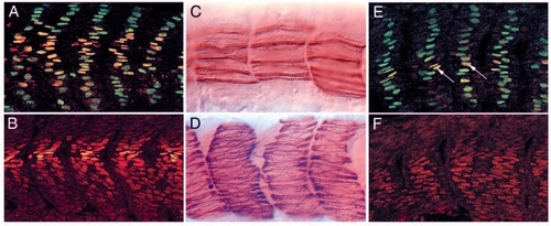
Cos2 is epistatic to shh and smo. (A) Eng expression in the MPs (long arrows) and MFFs (short arrow) revealed by histochemistry in a 24 hpf wild-type embryo. (B) Eng expression is absent from the myotome of a 24 hpf shh mutant embryo. (C) Restoration of Eng-expressing MP cells (arrows) in the myotome of a shh mutant embryo injected with cos2 MOs (n=9/12). (D) Myotome of a 24 hpf wild-type embryo stained with antibodies to slow MyHC showing the pattern of the SSFs. (E) Slow MyHC immunoreactivity is completely absent from the myotome of a 24 hpf smu mutant embryo, consistent with the lack all SSFs as well as MPs. Like syu embryos depicted in B, smu mutants also lack all Eng expression from the myotome (data not shown) (see also Barresi et al., 2000; Wolff et al., 2003). (F) Slow fibres are restored in significant numbers in smu mutants injected with cos2 MOs (n=15/21). |
Loss of Su(fu) activity enhances de-repression of Hh signalling in Cos2 morphants. (A) A 24 hpf wild-type embryo injected with MOs against Su(fu) and cos2 and stained with antibodies against Prox1 and Eng, showing very large numbers of supernumerary MP cells in the myotome (n=18/23). Compare with Fig. 4E,F. (B) The embryo depicted in A in medial view, showing extensive expansion in the numbers of Eng expressing MFFs (n=20/23). Compare with Fig. 4G,H. (C) Injection of Su(fu) MO into smu mutants restores some slow fibres (n=16/22). Compare with Fig. 6D,E. (D) Injection of Su(fu) as well as of cos2 MOs into smu embryos results in the recovery of substantial numbers of slow fibres (n=16/19). Compare with Fig. 6D,E. (E) A Su(fu) and cos2 MO co-injected smu embryo, stained with anti-Eng and anti-Prox1 antibodies, showing restoration of SSFs as well as MPs (arrows; n=10/18). (F) A Su(fu) and cos2 MO co-injected smu embryo, showing recovery of Eng expressing MFFs (n=16/18). |

Unillustrated author statements EXPRESSION / LABELING:
|