
- Title
-
Loss of gata1 but not gata2 converts erythropoiesis to myelopoiesis in zebrafish embryos
- Authors
- Galloway, J.L., Wingert, R.A., Thisse, C., Thisse, B., and Zon, L.I.
- Source
- Full text @ Dev. Cell

Loss of Gata1 Results in Expanded Myelopoiesis(A) pu.1 expression in control and gata1 MO-injected embryos at 14 somites (a, b), 18 somites (c, d), and 24 hpf (e, f). Embryos (a–d) were flat-mounted with anterior (left) and posterior (right). gata1 MO-injected embryos express pu.1 at 18 somites (d, brackets) and 24 hpf (f) while wild-type embryos have downregulated pu.1 transcripts (c, brackets, e) in the ICM precursors. pu.1 is also expressed in anterior myeloid precursors (a–f). gata1 MO-injected embryos have reduced expression of βe1 globin (h) in the ICM at 24 hpf compared to wild-types (g).(B) The number of cells expressing mpo and l-plastin are increased in the vessels of gata1 morphants (b, f, d, h) compared to wild-type embryos (a, e) and gata2 morphants (c, g). g1/g2 morphants (d, h) have decreased numbers of mpo- and l-plastin-expressing cells compared to the gata1 morphants alone (b, f).(C) Expression of c-myb (a–d) and ikaros (e–h) is maintained in the ICM cells of gata1, gata2, and g1/g2 morphants at 20 somites. pu.1 expression persists in the ICM of gata1 (j) and g1/g2 (l) morphants, but not gata2 morphants (k) or wild-type embryos (i). Expression of runx1 (m–p) and cebpα (q–t) persists in gata1 (n, r) and g1/g2 morphants (p, t) but not wild-type embryos (m, q) or gata2 morphants (o, s) at 22 hpf. cebpα is expressed at low levels in some ICM cells and in gut endoderm (q–t). ICM cells expressing mpo at 22 hpf are reduced in gata2 morphants (w) and lost in gata1 (v) and g1/g2 morphants (x) compared to wild-type embryos (u). Insets (u–x) are of cranial views of the same embryos showing normal mpo-expressing cells in wild-type (u) and gata1 morphants (v) and reduced numbers of mpo-expressing cells in gata2 (w) and g1/g2 morphants (x). EXPRESSION / LABELING:
|
Erythroid Precursors Are Converted into Myeloid Precursors in the Absence of Gata1(A) gata1-gfp cells from wild-type (a) and gata1 splice MO-injected (b) embryos at 10 somites (14 hpf) resemble hematopoietic precursors. At 24 hpf, most wild-type gata1-gfp cells resemble proerythroblasts (c, arrowheads), but myeloid cells with indented nuclei are observed. Cells from gata1 splice MO-injected embryos have myeloid features such as vacuoles and indented nuclei (d, arrowheads). Many erythrocytes and proerythroblasts are isolated from 48 hpf wild-type embryos (e, arrowheads) as well as a few myeloid cells. Cells from 48 hpf gata1 splice MO-injected embryos have vacuoles and indented nuclei (f, arrowheads), suggesting that they are myelomonocytes (scale bars are 10 μm).(B) Wild-type (a) and vlt mutant (c) embryos at 22 hpf have similar numbers of TUNEL-positive cells. vlt mutant embryos have an approximate 2-fold increase in apoptosis in their ICM region (d, inset) compared to their wild-type siblings (b, inset) at 28 hpf.(C) Confocal imaging of transverse sections of DAPI-stained (blue) embryos that have undergone double in situ hybridization for βe1 globin (green) and pu.1 (red). Wild-type embryos express βe1 globin and not pu.1 in their ICM cells at 22 hpf (a–c). gata1 MO-injected embryos express both βe1 globin and pu.1 in their ICM cells at 22 hpf (d–f). Some cells coexpress (yellow) both genes (d–f, arrowheads; n, notochord; nt, neural tube; scale bar equals 40 μm). EXPRESSION / LABELING:
|
Erythroid Genes Are Differentially Regulated by Gata1 and Gata2(A) Embryos (a–d) have been flat-mounted and photographed to show only the posterior tail region (anterior, left; posterior, right). Normal expression of gdpbp1 and epsin (a, c) is lost in ICM erythroid precursors of vlt mutants at 18 hpf (b, d). Expression of biliverdin reductase and SH3BP5 at 20 hpf (e) and 22 hpf (g), respectively, is absent in vlt mutant ICM cells (f, h).(B) In situ hybridization for βe1 globin (a–d), alas2 (e–h), carbonic anhydrase (i–l), and gata1 (m–p) at 20 hpf. Compared to wild-type embryos (a, e, i, m), ICM expression of βe1 globin, alas2, carbonic anhydrase, and gata1 was decreased in gata1 morphants (b, f, j, n) and absent in g1/g2 morphants (d, h, l, p). Decreased number of cells expressing βe1 globin, alas2, carbonic anhydrase, and gata1 are observed in gata2 morphants (c, g, k, o) compared to wild-types (a, e, i, m). EXPRESSION / LABELING:
|

Expression of Gata-Independent Erythroid Genes(A) Embryos were flat-mounted (anterior, left; posterior, right). Expression of biklf (a–d), KIAA0650 (e–h), testhymin (i–l), and kelch repeat-containing protein (m–p) is maintained in ICM precursors of 12 somite gata1, gata2, and g1/g2 morphants. Anterior expression of biklf stains the hatching gland (a–d).(B) ICM expression of biklf (a–d) at 20 hpf and KIAA0650 (e–h) and testhymin (i–l) at 22 hpf was decreased but present in gata1 morphants (b, f, j) and g1/g2 MO-injected embryos (d, h, l) compared to wild-type embryos (a, e, i). gata2 morphants (c, g, k) had decreased numbers of ICM cells expressing biklf, KIAA0650, and testhymin. EXPRESSION / LABELING:
|
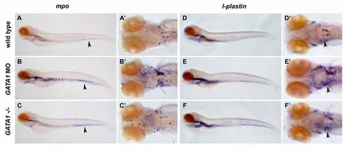
The gata1 MO-injected (B, B′) and vlt mutant embryos (C, C′) have an increased number of cells expressing mpo at 4 dpf compared to wild-types (A, A′). Expression of mpo in the tail (arrowheads) is especially affected. The number of cells expressing l-plastin at 4 dpf also increased in the gata1 MO-injected (E, E′) and vlt mutant embryos (F, F′) compared to wild-types (D, D′) particularly in the head regions (B′–F′). EXPRESSION / LABELING:
|

Wild-type gata1-egfp embryos have GFP in circulating blood cells in the vessels and over the yolk (A, arrowheads) and ectopically in the neural tube (asterisk). A gata1 morpholino targeting the exon/intron splice sites was used to avoid interference with transgene expression. At 48 hpf, Gata1-deficicent embryos had large, round, noncirculating GFP-positive cells distributed in a pattern resembling mpo expression (B). gata1 MO-injected gata1-egfp embryos express gfp in noncirculating cells in the major vessels and on the yolk (B, arrowheads). be1 globin (C) and mpo (E) are expressed normally in wild-type embryos. In gata1 MO-injected embryos, be1 globin expression is absent (D) and mpo expression is expanded (F). |
Reprinted from Developmental Cell, 8(1), Galloway, J.L., Wingert, R.A., Thisse, C., Thisse, B., and Zon, L.I., Loss of gata1 but not gata2 converts erythropoiesis to myelopoiesis in zebrafish embryos, 109-116, Copyright (2005) with permission from Elsevier. Full text @ Dev. Cell