
- Title
-
dlx3b and dlx4b function in the development of Rohon-Beard sensory neurons and trigeminal placode in the zebrafish neurula
- Authors
- Kaji, T., and Artinger, K.B.
- Source
- Full text @ Dev. Biol.
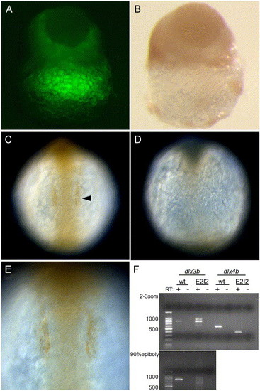
Knockdown of dlx proteins in embryos injected with dlx3b/dlx4b-ATG-MOs and dlx3b/dlx4b-E2I2-MOs. The knockdown by the ATG-MOs was revealed by immunostaining with anti-Dll antibody (A–E). (A and B) Late blastula embryos. (C–E) 6-somite-stage embryos. (C and E) Wild-type embryos. (A, B, D) ATG-MOs-injected embryos. (A and B) Same images of a blastula. (A) Fluorescence image revealing MOs + LFD, and (B) bright field. The dispersion of MOs and anti-Dll immunoreactivity are complementary in the epiblast. Lateral views. (C and D) Anti-Dll antibody immunoreactivity in the otic placodal primordium (arrowhead) is absent in MOs-injected embryos. Dorsal views, anterior is to the top. (E) The magnification view of the otic placodal primordium in (C). The knockdown by the E2I2-MOs was revealed by RT-PCR followed by gel electrophoresis (F). (F) Wild type (wt) as compared to E2I2 MO (E2I2) dlx3b and dlx4b RT-PCR products. In dlx3b RT-PCR, at 2–3-somite stage, the amplification of cDNA from wild-type embryos produced an 810-bp band, and cDNA from E2I2-MOs-injected embryos produced three bands of 630, 810, and 900-bp in size. At 90% epiboly, in E2I2-MOs-injected embryos, the 810-bp band was faint. In dlx4b RT-PCR, at 2- to 3-somite stage, the amplification of cDNA from wild-type embryos produced a 580-bp band, and cDNA from E2I2-MO-injected embryos produced a 390-bp band. Negative-control reactions run in parallel, where reverse transcriptase was omitted in the cDNA synthesis reactions (RT-). |

Phenotypes of Rohon-Beard (RB) sensory neurons in MOs and EnR-dlx3bhd mRNA injected embryos at 3- and 6-somite stage. (A–J) Embryos at 3-somite stage. Dorsal views, anterior is to the top. (K–P) Embryos at 6-somite stage. (A, F, K, M, O) Wild-type embryos. (B, G, L, N, P) 20 ng ATG-dlx3b-MO/20 ng ATG-dlx4b-MO-injected embryos. (C and H) 20 ng E2I2-dlx3b-MO/20 ng E2I2-dlx4b-MO-injected embryos. (D and I) One hundred fifty-picogram EnR-dlx3bhd mRNA-injected embryos. (E and J) Forty-nanogram control MO-injected embryos. (A–E, M–P) Expression of HuC. (F–J) Expression of tlx3a. (K and L) Expression of isl-1. (O) High magnification lateral view of a wild-type embryo. (P) High magnification lateral view of (N). In (A), RB neurons (rb) and primary motor neurons (arrowhead) are seen. After injection of dlx3b and dlx4b-MOs or EnR-dlx3bhd mRNA (B–D), primary motor neurons are present, but RB neurons are highly reduced or absent, as seen by the expression of HuC. A control MO injected at the same concentration in (E) has normal RB neurons. In (G–I), RB neurons are highly reduced or absent after injection of dlx3b and dlx4b-MOs or EnR-dlx3bhd mRNA, observed with tlx3a expression. In a control MO injected at the same concentration in (J), no changes are observed. In embryos allowed to develop until the 6-somite stage (L and N), RB neurons continue to show a reduction after injection of dlx3b and dlx4b-MOs. The bracket in (L) shows the region of no RB neurons. In a higher magnification view (O and P), it is easily recognized that RB neurons are highly reduced in MO-injected embryos observed at 6-somite stage. EXPRESSION / LABELING:
PHENOTYPE:
|

Phenotypes of neurons in the trigeminal placodes in MOs and EnR-dlx3bhd mRNA injected embryos at 3- and 6-somite stage. (A–J) Embryos at 3-somite stage. Anterior views. (K–N) Embryos at 6-somite stage. Anterior views. (A, F, K, M) Wild-type embryos. (B, G, L, N) 20 ng ATG-dlx3b-MO/20 ng ATG-dlx4b-MO-injected embryos. (C and H) 20 ng E2I2-dlx3b-MO/20 ng E2I2-dlx4b-MO-injected embryos. (D and I) One hundred fifty-picogram EnR-dlx3bhd mRNA-injected embryos. (E and J) Forty-nanogram control MO-injected embryos. (A–E, K, L) Expression of HuC. (F–J) Expression of neuroD/pax2a. (M and N) Expression of isl-1. In a wild-type embryo (A), neurons in the trigeminal placodes (tg) and anterior lateral/medial hindbrain interneurons (li, mi) are observed. After injection of dlx3b and dlx4b-MOs or EnR-dlx3bhd mRNA shown in (B–D), anterior lateral/medial hindbrain interneurons remain, but neurons in the trigeminal placodes are highly reduced or absent as shown by expression of HuC. In a control-MO injected embryo (E), neurons in the trigeminal placodes are seen in the placodal primordial region. neuroD/pax2a expression in (G–I), both are highly reduced or absent in the placodal primordial region following injection of dlx3b and dlx4b-MOs or EnR-dlx3bhd mRNA. Midbrain–hindbrain boundary is seen in the central nervous system (mhb). In a control MO-injected embryos (J), no changes were observed in the trigeminal (tg) and otic placodal primordia (ot). Shown at the 6-somite stage, expression of HuC (L) and isl-1 (N), neurons in the trigeminal placodes remain highly reduced, as compared to wild-type embryos in (K and M), respectively. EXPRESSION / LABELING:
PHENOTYPE:
|

HuC expression in embryos injected with dlx3b-ATG-MO and dlx4b-ATG-MO and EnR-dlx3bhd mRNA at 3-somite stage. (A, C, E, G, I) Dorsal views, anterior is to the top. (B, D, F, H, J) Anterior views. (A and B) Wild-type embryos. (C and D) 8 ng dlx3b-MO- and 8 ng dlx4b-MO-injected embryos. (E and F) 16 ng dlx3b-MO- and 16 ng dlx4b-MO-injected embryos. (G and H) 60 pg EnR-dlx3bhd mRNA-injected embryos. (I and J) 120 pg EnR-dlx3bhd mRNA-injected embryos. (A–F) and (G–J) show that the expression of HuC, which is expressed in RB neurons in the trunk (rb) and neurons in trigeminal placodes in the head (tg), is gradually reduced as the concentration of MOs and EnR-dlx3bhd mRNA are increased. In contrast, primary motor neurons (arrowhead) and anterior lateral hindbrain interneurons (li) and anterior medial hindbrain interneurons (mi) are not changed. EXPRESSION / LABELING:
PHENOTYPE:
|
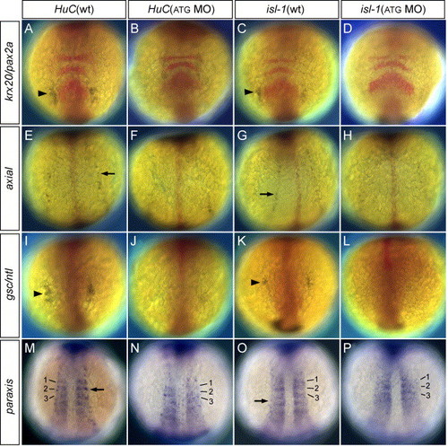
Double in situ hybridization of HuC/isl-1 and neural/mesodermal markers in MOs-injected embryos. (A, C, E, G, I, K, M, O) Wild-type embryos at 3-somite stage. (B, D, F, H, J, L, N, P) 20 ng ATG-dlx3b-MO/20 ng ATG-dlx4b-MO-injected embryos at 3-somite stage. (A and B) Expression of HuC (blue) and krx20/pax2a, a marker of rhombomeres 3 and 5 and midbrain–hindbrain boundary (red). (C and D) Expression of isl-1 (blue) and krx20/pax2a (red). (E and F) Expression of HuC (blue) and axial marking floor plate (red). (G and H) Expression of isl-1 (blue) and axial (red). (I and J) Expression of HuC (blue) and gsc/ntl expressed in the prechordal plate and notochord (red). (K and L) Expression of isl-1 (blue) and gsc/ntl (red). (M and N) Expression of HuC (blue) and paraxis showing expression in the somites (blue). Numbers 1–3 correspond to the somites. (O and P) Expression of isl-1 (blue) and paraxis (blue). Numbers 1–3 correspond to the somites. In (B, D, J, L), neurons in the trigeminal placodes (arrowhead) are highly reduced or absent, shown by the expression of HuC and isl-1. The expression of HuC and isl-1 in the RB neurons (F, H, N, P; arrow) are highly reduced or absent. However, no changes are observed in neural/mesodermal marker expression in dlx3b and dlx4b MOs-injected embryos (B, D, F, H, J, L, N, P). EXPRESSION / LABELING:
PHENOTYPE:
|
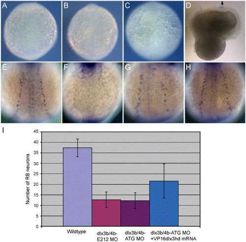
Study of necrosis and rescued phenotypes of RB neurons by VP16-dlx3bhd mRNA in dlx3b and dlx4b MOs-injected embryos. Trypan blue-stained pattern in 2-3-somite-stage embryos (A–D) and HuC-expressing RB neuron pattern in 3-somite-stage embryos (E–H). (A) A wild-type embryo. (B) A 20-ng ATG-dlx3b-MO/20 ng ATG-dlx4b-MO-injected embryo. (C) A 40-ng control MO-injected embryo. (D) An embryo injured by a tungsten needle. No trypan blue-stained pattern is seen in wild type, 20 ng ATG-dlx3b-MO/20 ng ATG-dlx4b-MO-injected, and 40 ng control MO-injected embryos (A–C). In the injured embryo (D), a blue-stained pattern can be seen (arrow), indicating cell necrosis. (E) A wild-type embryo expressing HuC. (F) An embryo injected with 20 ng ATG-dlx3b-MO/20 ng ATG-dlx4b-MO. (G) A partially rescued embryo in RB neurons by VP16-dlx3bhd mRNA, injected with 20 ng ATG-dlx3b-MO/20 ng ATG-dlx4b-MO. (H) A fully rescued embryo in RB neurons by VP16-dlx3bhd mRNA, injected with 20 ng ATG-dlx3b-MO/20 ng ATG-dlx4b-MO. (I) Numbers of RB neurons are recovered in MO-injected embryos by injection of 150 pg VP16-dlx3bhd mRNA. EXPRESSION / LABELING:
|

Expression of the upstream gene, neurogenin1, for HuC, tlx3a, and neuroD marking RB neurons and neurons in the trigeminal placodes, in embryos injected with dlx3b-MO and dlx4b-MO at 3-somite stage. (A and C) Wild-type embryos. (B and D) 20 ng ATG-dlx3b-MO/20 ng ATG-dlx4b-MO-injected embryos. (A and B) The expression of neurogenin1 in RB neuron precursors (rb) is normal in MO-injected embryos. Dorsal views. (C and D) The expression of neurogenin1 in the precursors of neurons in the trigeminal placodes (tg) is absent in MOs-injected embryos (D). Anterior views. mn, primary motor neuron precursors; in, primary interneuron precursors; mi, anterior medial hindbrain interneuron precursors; li, anterior lateral hindbrain interneuron precursors. EXPRESSION / LABELING:
|

Expression of bmp2b, 4, and 7 in embryos injected with dlx3b-MO and dlx4b-MO at 90% epiboly. Expression in wild-type embryos of bmp2b (A), bmp4 (C), and bmp7 (E). Expression in 20 ng ATG-dlx3b-MO/20 ng ATG-dlx4b-MO-injected embryos of bmp2b (B), bmp4 (D), and bmp7 (F). (A and B) show that there is a strong triangle of bmp2b expression (arrowhead) at the border of the non-neural ectoderm in the wild-type embryos but this region is lost in MOs-injected embryos. Ventral bmp2b expression persisted in MOs-injected embryos (arrow). No alterations are seen in the expression of bmp4 and bmp7 between wild-type embryos and MOs-injected embryos (C–F). Lateral views, and dorsal is to the right. EXPRESSION / LABELING:
|

HuC expression in mosaic embryos between wild-type and MOs-injected embryos. (A–C) Wild-type embryos at tailbud stage. Lateral views, and dorsal is to the right. (D–O) Transplanted embryos at 3-somite stage. D–I are the wild-type host embryos. (J–O) are the MOs-injected host embryos. (D and J) Dorsal views. (G and M) Anterior views. (A) Expression pattern of dlx3b (arrowheads). (B) Expression pattern of neurogenin1 (double arrowhead in the trunk and double arrow in the head). (C) Expression pattern of dlx3b and neurogenin1. (D–O) HuC expression in mosaic embryos. (A–C) show that dlx3b expression (arrowheads) and neurogenin1 expression in the lateral trunk (double arrowhead) are adjoining but not overlapping in the trunk. In contrast, dlx3b expression and neurogenin1 expression in the lateral head (double arrow) are overlapping. D–F show that RB neurons (rb) do not develop in the neural plate where brown-labeled MOs-injected cells (mo) are in a narrow region (bracket) just outside the neural plate. dlx3b and dlx4b should be expressed in this narrow region under wild-type conditions. (E) A high magnification view of RB neurons (rb) on the left side of embryo in D. (F) A high magnification view of the narrow region defined by brackets on the right side of embryo in D. The high number of MO-injected cells (brown, mo) is able to inhibit HuC expression in neighboring cells. G–I show that neurons in the trigeminal placodes (tg) do not develop there when brown-labeled MOs-injected cells occupy the trigeminal placode (circle). dlx3b and dlx4b are expressed in this circle region under wild-type conditions. (H) A high magnification view of neurons in the trigeminal placode (tg) on the right side of embryo in G. (I) A high magnification view of the trigeminal placodal region defined by the circle on the left side of embryo in G. The high number of MO injected cells (brown, mo) is able to inhibit HuC expression in the trigeminal placode. (J–L) In a MO-injected background, RB neurons are seen only around the brown-labeled wild-type cells (wt) in the embryonic epidermis. The host MOs-injected embryos develop no or reduced levels of RB neurons. (K) A high magnification view of RB neurons on the left side of embryo in J. RB neurons can be seen only around the brown wild-type cells (wt). (L) A high magnification view of RB neurons on the right side of embryo in J. RB neurons can be seen only around the brown wild-type cells (wt). (M–O) Neurons in the trigeminal placodes develop when brown-labeled wild-type cells occupy the trigeminal placode (square) in the host MOs-injected embryos having no trigeminal placodal neurons. N is the high magnification view of the trigeminal placode defined by the square on the right side of embryo in M. The high number of wild-type cells expresses HuC in themselves, so many double-labeled neurons can be seen in the trigeminal placode (arrow). (O) A high magnification view of the trigeminal placodal region on the left side of embryo in M. No neurons in the trigeminal placode are observed where wild-type cells are not present. li, anterior lateral hindbrain interneuron; mi, anterior medial hindbrain interneuron. All scale bars, 50 μm. Scale bar in A applies for A, B, and C. Scale bar in F applies for both E and F, etc. EXPRESSION / LABELING:
|
Reprinted from Developmental Biology, 276(2), Kaji, T., and Artinger, K.B., dlx3b and dlx4b function in the development of Rohon-Beard sensory neurons and trigeminal placode in the zebrafish neurula, 523-540, Copyright (2004) with permission from Elsevier. Full text @ Dev. Biol.