
- Title
-
A moving wave patterns the cone photoreceptor mosaic array in the zebrafish retina
- Authors
- Raymond, P.A., and Barthel, L.K.
- Source
- Full text @ Int. J. Dev. Biol.
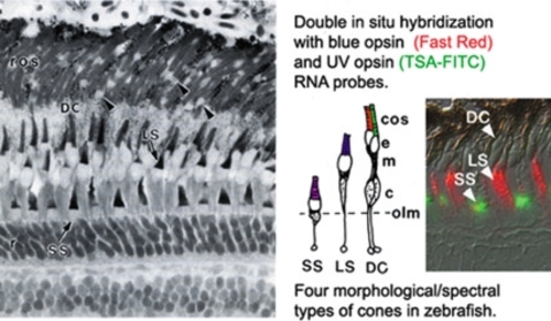
Spectral subtypes of photoreceptors in zebrafish. Radial, histological section of adult zebrafish retina, with the retinal pigmented epithelium and choroid at the top. Rod nuclei (r) in the outer nuclear layer, are connected by thin myoid processes to ellipsoids (arrowheads) and rod outer segments (ros). The four morphological/spectral cone subtypes are shown in the cartoon, with the cone outer segments (cos) color-coded to indicate the spectral peak of the opsin protein expressed in short single (SS), long single (LS) and double cones (DC). Other abbreviations: ellipsoid, e; myoid, m; cone nucleus, c; outer limiting membrane (olm) of the retina. The insert shows double in situ hybridization with two different opsin RNA probes and fluorescent detection. Methods: RNA probes are tagged with digoxigenin (DIG) or fluorescein (FL), respectively, and are detected with anti-DIG and anti-FL antibodies conjugated to horseradish peroxidase (HRP) or alkaline phosphatase (AP), respectively, and visualized with enzymatic substrates that produce contrasting Fig. 2. Cone mosaic pattern in zebrafish. En face views of isolated, flattened, adult zebrafish retinas, processed for in situ hybridization with cone opsin RNA probes (fluorescent signals are pseudo-colored to correspond to the spectral subtype). At lower left is a cartoon schematic of the cone mosaic pattern. fluorescent precipitates (Barthel and Raymond, 2000; Jowett, 2001). The Fast Red AP substrate is bright red in visible light, and it also fluoresces red with a rhodamine filter set. The TSA Biotin System uses HRP to catalyze the deposition of biotin-labeled tyramide, which is recognized by a streptavidin-fluorophore conjugate, such as fluorescein isothiocyanate (FITC). We performed control experiments to verify that the different detection methods have comparable sensitivity. |

Cone mosaic pattern in zebrafish. En face views of isolated, flattened, adult zebrafish retinas, processed for in situ hybridization with cone opsin RNA probes (fluorescent signals are pseudo-colored to correspond to the spectral subtype). At lower left is a cartoon schematic of the cone mosaic pattern. |
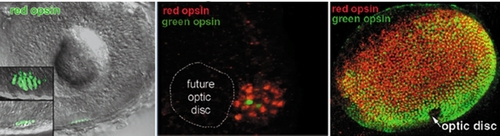
Development of the cone mosaic pattern. Laser scanning confocal images (3D projections) from whole eyes of zebrafish embryos, processed for in situ hybridization with red and green opsin RNA probes to visualize individual cone photoreceptors. After processing, whole eyes are isolated from the embryonic heads and visualized using a Zeiss 510 laser scanning confocal microscope, by collecting a set of optical sections through the entire thickness of the eye (~100 µm). Computer reconstructions of three-dimensional and projection images were then generated from various angles of view. (A) The earliest photoreceptors appear at ~2 days postfertilization (dpf) in the outer nuclear layer of the ventral-nasal retina adjacent to the optic stalk, and the first cone opsin is red (TSA method, FITC green fluorophore). The lens is visible in the center, and the inset shows the individual red cones at higher magnification, in orthogonal views. Note the regularly spaced rows of red cones in the rotated (planar) view. (B) Double in situ hybridization with red opsin (Fast Red) and green opsin (TSA-FITC) probes, showing (at high magnification) the ventral patch of initial cones, adjacent to the future optic disc. (C) By ~3 dpf, the front of the wave of differentiating photoreceptors has nearly reached the dorsal-temporal edge of the retina at the upper left corner, with red cones at the leading edge, trailed by green cones. EXPRESSION / LABELING:
|
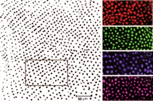
Computational analysis of cone mosaic patterns. (A) Scion Image data file of an adult retinal whole mount processed for in situ hybridization (blue opsin), in which the area selected for analysis is boxed. (B-E) Confocal images of embryonic retinas at ~3dpf processed with red, green, blue or UV opsin probe (pseudo-colored), respectively. EXPRESSION / LABELING:
|