
- Title
-
Inhibition of Wnt/Axin/{beta}-catenin pathway activity promotes ventral CNS midline tissue to adopt hypothalamic rather than floorplate identity
- Authors
- Kapsimali, M., Caneparo, L., Houart, C., and Wilson, S.W.
- Source
- Full text @ Development

mbl embryos show altered AP regionalisation of the ventral midline of the neural plate. Dorsal views of wild-type and mbl embryos with anterior on the top. In this and other figures, where indicated, stage is shown bottom left, genes analysed by in situ hybridization are shown bottom right and the experimental procedure is top right. Arrowheads point to hypothalamic (nk2.1a) gene expression. pax2.1 is used as a marker of the prospective midbrain in A-H and K-L. The asterisk indicates the prospective hypothalamic domain of reduced foxa2 expression in a wild-type embryo (C) and brackets indicate the AP length of the prospective floorplate domain rostral to the midbrain in wild-type (C,G) and mbl (D,H) embryos. The inset panel between G and H shows nk2.1a expression in the prospective hypothalamus in yellow and foxa2 in the prospective floorplate in blue. The margin of the neural plate, the hatching gland, the prospective notochord and eye field (red) are shown by dlx3, hgg1, ntl and rx3 expression domains in I,J. The mbl embryo lacks rx3 expression in the eye field. (K-L) Transplanted cells overexpressing wnt8b (brown) suppress nk2.1a expression (blue, arrowhead) and expand rostrally pax2.1 expression (white asterisk) in a wild-type host embryo. Abbreviations: tb, bud stage, e, prospective eye, hg, hgg1 expression in the prechordal plate, hy, hypothalamus, mb, midbrain, n, prospective notochord; s, somite stage; wt, wild type |

mbl embryos show a reduction in the size of the hypothalamus coupled with an expansion in posterior diencephalic/midbrain floorplate. Lateral views of brains of embryos between 25 and 31 somites with anterior to the left. (A-F) The mbl embryos (B,D,F) show reduced hypothalamic tissue and rostral expansion of floorplate-expressed genes compared to the wild-type embryos (A,C,E). Arrowheads point at the rostral limit of floorplate marker expression (A-D). Brackets show the AP expansion of diencephalic/midbrain foxa1 expression (red) in the mbl (F) compared to the wild-type embryo (E). (G-H) Transplanted cells overexpressing axin1 (brown) restore nk2.1a expression (blue, asterisk) in the rostral ventral neural tube of an mbl embryo. Abbreviations: hy, hypothalamus; mfp, medial floorplate; t, telencephalon; zli, zona limitans intrathalamica; wt, wild type. |
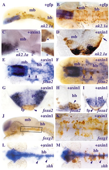
Exogenous Axin1 promotes hypothalamic and suppresses floorplate marker gene expression. Lateral (A,C,J) and dorsal (B,E,F,K,L,M) views and 14 μm transverse sections (D,G-I) of brains of embryos between 22 and 28 somites in which cells (brown) expressing various constructs (top right) were transplanted into the prospective hypothalamus/anterior floorplate of 60-65% epiboly stage hosts. Sections are at the level of the ventral midbrain (D,G,I) and the hindbrain (H). (A-B) gfp-expressing cells (brown) integrate in the ventral midbrain and hindbrain but do not express nk2.1a. (C-D) Cells overexpressing axin1 (brown, asterisks in the insets) in the midbrain and hindbrain of two different embryos ectopically express nk2.1a (blue, asterisks). The arrow points to the absence of ectopic nk2.1a expression in more dorsal cells overexpressing axin1 (brown) in the midbrain. (E-I) Floorplate marker expression (blue) is downregulated in axin1 overexpressing cells (brown, arrowheads). The expression of foxa2 is shown in the same embryo after in situ hybridization (E) and after immunohistochemistry to reveal the axin1 overexpressing cells (brown, F) and in transverse sections (G,H). The arrowheads point to sites of downregulation of foxa2 or foxa1 in the transplanted cells in the midline of the midbrain (E-G,I) and hindbrain (H). (J-K) axin1 overexpressing cells do not ectopically express foxg1 in the posterior ventral neural tube of the embryo (J) and in higher magnification (K). (L-M) Exogenous axin1 expands shh expression (arrowheads) lateral to the CNS midline shown after in situ hybridization (L) and after immunohistochemistry to reveal the transplanted cells (M). Abbreviations: mb, midbrain; hb, hindbrain; fp, floorplate; pd, posterior diencephalon.
|

Suppression of Wnt signalling in combination with activation of Nodal signalling promotes hypothalamic marker gene expression. Lateral (A,B,C,E,F) and ventral (D) views of brains of embryos at 24-somite stage with anterior to the left in which cells (brown) overexpressing hdl/tcf3a (A,B), axin1 (C), dkk1 (D), madh2CA (E) or madh2CA+dkk1 (F) were transplanted into the prospective hypothalamus/anterior floorplate at 60-65% epiboly. A and B show the same embryo before and after immunohistochemistry for Gfp. In A,B,C,F, black arrowheads point to ectopic nk2.1a expression in the medial floorplate and/or adjacent cells. In B,C,E, red arrows point to cells that integrated in the medial floorplate. In D, Dkk1+ cells (brown) mainly integrate in more lateral positions in the neuroepithelium compared to the Axin1+ and Madh2CA+ cells that integrate in the medial floorplate (brown, red arrows) of the embryos in C and E respectively. Abbreviations: hy, hypothalamus; mb, midbrain; mfp, medial floorplate; pv, posterior-ventral hypothalamus. |

mbl embryos lose rostral but retain caudal hypothalamic marker gene expression. (A-H) Lateral views of brains of embryos at about 31 somites with anterior to the left. (A-D) The hypothalami of the mbl embryos (B,D) do not express rx3 (B) and lose the anterior domain of shh expression (D) compared to the wild-type embryos (A,C). (E-F) The mbl embryo (F) retains expression of the hypothalamic marker emx2 which is expressed in the caudal hypothalamus of the wild-type embryo (E). Arrowheads point at the caudal limit of the hypothalamus. (G-H) Transplanted cells overexpressing axin1 (brown) that have incorporated into the rostral ventral CNS restore rx3 expression (blue) in the mbl embryo (G). (H) A higher magnification view of the transplant (boxed) of this embryo. (I-J) Some Axin1-overexpressing cells that incorporate into the floorplate domain of a wild-type host ectopically express emx2 (arrows). The transverse section (indicated by the left arrow in I) shows ectopic emx2 expression in the floorplate (arrowhead in J). Abbreviations: ad, anterior-dorsal hypothalamus; fp, floorplate; mfp, medial floorplate; pv, posterior-ventral hypothalamus; t, telencephalon; zli, zona limitans intrathalamica. |
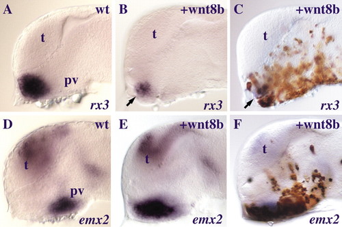
wnt8b overexpression promotes expression of posterior hypothalamic markers. Lateral (A-F) views of brains of wild-type embryos (A,D) and embryos in which cells overexpressing wnt8b (B,C,E,F) have incorporated into the hypothalamus. C and F show the same embryos as B and E after immunocytochemistry to reveal the positions of cells expressing wnt8b. The arrows in B,C point to decreased rx3 expression (B,C) compared to control (A). Abbreviations: pv, posterior-ventral hypothalamus; t, telencephalon. |