
- Title
-
Fgf signaling induces posterior neuroectoderm independently of Bmp signaling inhibition
- Authors
- Rentzsch, F., Bakkers, J., Kramer, C., and Hammerschmidt, M.
- Source
- Full text @ Dev. Dyn.
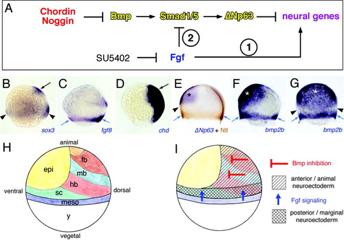
Diagram illustrating the epistatic relationships between Chordin and Noggin, bone morphogenetic proteins (Bmps), Smad transcription factors, and ΔNp63 during neural induction. In addition, two alternative routes of fibroblast growth factor (Fgf) function are indicated, acting either by means of an inhibition of Smad proteins (2) or independently of Bmp signaling (1). B-G: Expression pattern of genes indicated in the lower right corner at midgastrula stages (80%-90% epiboly). B-F show the lateral view, dorsal to the right, anterior/animal pole up; and G shows the ventral view. Blue arrows indicate mesoderm of germ ring, black arrows anterior/animal neuroectoderm, arrowheads posterior/marginal neuroectoderm, and stars non-neural ectoderm. H: Distribution of ectodermal and mesodermal cells in midgastrula embryo, modified from Woo and Fraser (1995) and Thisse et al. (2000). I: Diagram illustrating differential induction of anterior neuroectoderm by Bmp inhibitors from the dorsal axis and of posterior neuroectoderm by Fgf signaling from the germ ring mesoderm. chd, chordin; Ntl, No tail (Brachyury/T); fb, forebrain; mb, midbrain; hb, hindbrain; sc, spinal cord; meso, mesoderm; y, yolk. EXPRESSION / LABELING:
|
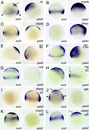
Differential effects of gain or loss of fibroblast growth factor (Fgf), bone morphogenetic protein (Bmp), or ΔNp63 function on ectodermal patterning. Panels show in situ hybridizations for sox3 (neuroectoderm) on the left and for gata2 (non-neural ectoderm) on the right, 80% epiboly, dorsal to the right, animal/anterior pole up. A,J: Arrows indicate sox3 expression in anterior neuroectoderm, arrowheads sox3 expression in posterior/marginal neuroectoderm. Injected mRNAs and SU5402 treatments (SU) are indicated in the upper right corner. E,H,I: Embryos were treated with 20 μM from 4 hpf to 8 hpf, embryos in F with 20 μM SU5402 from 5.5 hpf to 8 hpf. H: Coinjection of noggin mRNA abolishes the endogenous but not the SU-induced ectopic marginal gata2 expression (indicated with an arrow). L: Coinjected ΔNp63 mRNA abolishes anterior sox3 expression, and rescues gata2 expression (indicated with an arrow), whereas it cannot block the Fgf-induced expansion of the marginal/posterior sox3 domain. Siblings of embryos shown in all panels were also stained for the anterior neural marker otx2 (Li et al., 1994) and the posterior neural marker hoxb1b (Alexandre et al., 1992), leading to consistent results (Fig. 3, and data not shown). ctr, uninjected or untreated control embryo. EXPRESSION / LABELING:
|

Differential effects of loss of fibroblast growth factor (Fgf) signaling and gain of bone morphogenetic protein (Bmp) or ΔNp63 function on hoxb1b and n-cadherin expression. A-D: In situ hybridizations for hoxb1b (posterior neuroectoderm) at midgastrula stage (80% epiboly), lateral views, dorsal to the right. E-H: In situ hybridizations for ncadherin (ncad), late segmentation stage (24 hours postfertilization), lateral views, anterior to the left, dorsal up. Injected mRNAs and SU5402 treatments (SU) are indicated in upper right corner. E: Arrow indicates ncad-positive cells in the spinal cord of the trunk region. G: bmp2b mRNA-injected embryo displays strongest ventralization (V5; onion-like morphology; Kishimoto et al., 1997) EXPRESSION / LABELING:
|
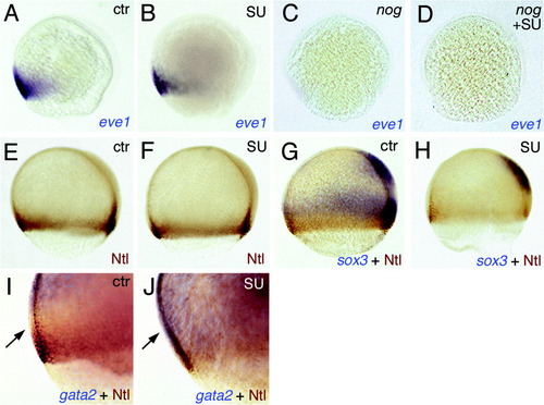
SU5402-treated embryos display normal expression of mesodermal marker genes. All embryos are shown at midgastrula stages (80% epiboly), lateral views, dorsal to the right. In situ probes (in blue) and antibodies (in brown) used are indicated in lower right corner. I,J: The arrow in I indicates the stripe between the gata2-positive non-neural ectoderm and the Ntl-positive mesoderm, which accommodates the posterior-most neuroectoderm and which has become gata2-positive in the SU5402-treated embryo (J). EXPRESSION / LABELING:
|