
- Title
-
Zebrafish mnx genes in endocrine and exocrine pancreas formation
- Authors
- Wendik, B., Maier, E., and Meyer, D.
- Source
- Full text @ Dev. Biol.

Expression of zebrafish mnx genes in early embryogenesis. Expression of mnr2a (A–E), mnr2b (F–J) and hb9 (K–O) in whole embryos at tailbud stage (A, F, K), two somite stage (B, G, L), 10 somite stage (C, H, M) and at 20 somite stage (D, I, N). (E, J, O) Higher magnification of the tail region of 20-somite stage embryos. Embryos are shown from dorsoposterior with anterior up (A–C, F–H, K–M) or from lateral with dorsal up and anterior to the left (D, E, I, J, N, O). Indicated are expression domains in the ventral spinal cord (black arrowhead), lateral positioned mesoderm (white arrowhead), endoderm cells (arrow), notochord (n), hypochord (hc), ventral tail mesoderm (tm) and tailbud (asterisk). Note the complementary axial expression of hb9 and mnr2b in notochord and tail bud. EXPRESSION / LABELING:
|
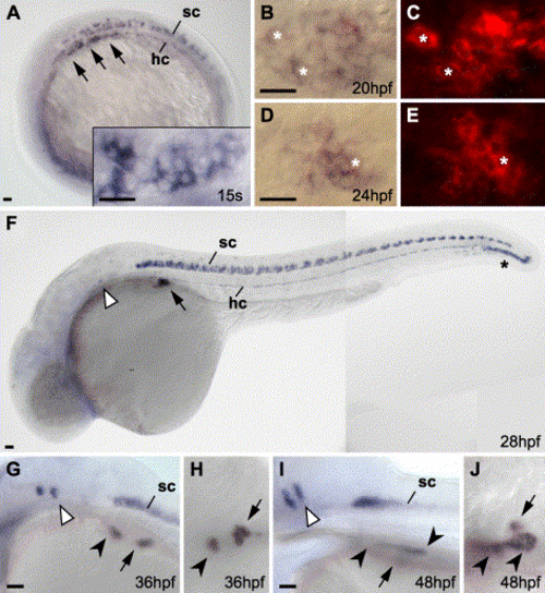
Tissue-specific expression of hb9 in endocrine pancreas. Expression of hb9 in purple at 15 somite (A), 20 hpf (B, C), 24 hpf (D, E), 28 hpf (F), 36 hpf (G, H) and 48 hpf (I, J). Between 15 somite stage (inlay in A shows a dorsal view of the same embryo in a higher magnification) and 24 hpf endodermal hb9 expression changes from an elongated mono-layered domain to a condensed structure underlying the hypochord (hc). (B–E) Bright field (B, D) and fluorescence (C, E) images of double in situ stains for hb9 (NBT/BCIP) and insulin (Fast Red) show expression of insulin in most hb9-expressing cells; asterisks mark examples for double-labeled cells. Further indicated are hb9 expression domains in the spinal cord (sc), rhombomeres 5 and 6 (white arrowheads) and the swim bladder (black arrowheads). Embryos are shown from a lateral (A, F, G, I) or dorsal view (B, C, D, E, H, J) with anterior to the left. Scale bars correspond to 20 μm. EXPRESSION / LABELING:
|

Expression of mnr2a in the exocrine pancreas. (A–C) Expression of mnr2a at 25 hpf (A), 36 hpf (B) and 96 hpf (C). Indicated are mnr2a expression domains in the endoderm (black arrowhead), in rhombomeres 5, 6 (white arrowhead) and in the spinal cord (sc). (D–J) Double labeling of mnr2a (purple) and insulin (red) expression at 25 hpf (D, E), 33 hpf (F), 36 hpf (G), 48 hpf (H), 70 hpf (I) and 84 hpf (J). Arrows in E, F and G mark mnr2a-positive close to insulin-expressing cells, note that mnr2a and insulin are expressed in different cells; the bar marks the position of the notochord (n). (K) Overlapping expression of mnr2a (blue) and trypsin (red) at 96 hpf. Embryos are shown from lateral (A–C, E) or dorsal (D, F–K) with anterior to the left. Scale bars correspond to 50 μm. |
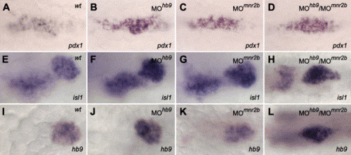
mnr2b and hb9 are not required for the initiation of pancreas formation. Similar expression of pdx1 at 20 hpf (A–D), and isl1 (E–H) and hb9 (I–L) at 36 hpf in wild type embryos (A, E, I) and in embryos injected with 4 ng MOhb9 (B, F, J) or 4 ng MOmnr2b (C, G, K) or 4 ng MOhb9 and 4 ng MOmnr2b (D, H, L) (unchanged expression levels were found for A: n = 15/15, B: n = 16/16, C: n = 19/19, D: n = 15/16, E: n = 11/11, F: n = 9/11, G: n = 13/13, H: n = 10/13, I: n = 20/20, J: n = 12/15, K: n = 12/12, L: n = 21/23). Slightly reduced expression levels were found in few hb9 morphants (F: n = 2/11, J: n = 3/15) and hb9-mnr2b double morphants (D: n = 1/16, H: n = 3/13, L: n = 2/23). Notably, 2 of the uninjected embryos and 3 hb9 morphants showed split pdx1 expression, while expression levels were not changed. All embryos are shown from the ventral with anterior to the right. Expression of isl1 (E–H) marks prospective endocrine pancreas cells (right expression domain) and a population of cells that remains to be determined (left expression domain). Also note that the expression levels of pdx1, isl1 (right domain only) and hb9 are similar in morphants and wild types, while the endocrine expression domains of isl1 and hb9 are more condensed and extended along the anterior–posterior axis in hb9/mnr2b double morphants (H, L) than in the wild types (E, I) and single morphants (F, G, J, K). Scale bars correspond to 50 μm. EXPRESSION / LABELING:
PHENOTYPE:
|
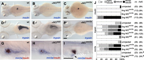
hb9 and mnr2a are differentially required in endocrine and exocrine pancreas formation. Expression of insulin at 3.5 dpf (A–C), trypsin at 4 dpf (D–F), and mnr2a (purple) and insulin (red) at 3.5 dpf in higher magnification (G–I) in wild type embryos (A, D, G) and in embryos injected with 2 ng MOhb9 (B, E, H) or 4 ng MOmnr2a (C, F, I). (G–I) Expression of insulin is strongly reduced in embryos injected with MOhb9 (B, H) but not in embryos injection of MOmnr2a (C, I). In contrast, expression of trypsin is strongly reduced after injection of MOmnr2a (F) but is not changed after injection of MOhb9 (E). Similarly, mnr2a expression is only reduced in MOmnr2a-injected embryos but not in MOhb9-injected embryos (I); arrow marks mnr2a expression. Embryos are shown from dorsal (A–F) or in a higher magnification from lateral with anterior left (G–I). (J) Summary of morpholino experiments. Indicated numbers of embryos (n) were injected with the indicated type and amount of morpholino and analyzed for expression of insulin, trypsin or mnr2a at indicated stages (hpf). The phenotypes of the embryos were classified relative to the expression level of the marker genes in the un-injected embryos: average wild type level (wt), reduced up to 50% (slightly reduced), reduction up to 10% (reduced), reduced to less than 10% or missing (strongly reduced). While injection of MOhb9 or MOmnr2a could result in total loss of insulin or trypsin expression, respectively, we did not find MOmnr2a embryos entirely lacking endoderm mnr2a expression. Note that the un-injected embryos frequently show different levels of mnr2a and trypsin expression representing natural variations found in zebrafish. (#) All analyzed embryos showed similar level of expression and since only few mnr2a-positive cells can be detected at 54 hpf, the presence of mnr2a-expressing cells was classified as 100%. Scale bars correspond to 100 μm. EXPRESSION / LABELING:
PHENOTYPE:
|
Reprinted from Developmental Biology, 268(2), Wendik, B., Maier, E., and Meyer, D., Zebrafish mnx genes in endocrine and exocrine pancreas formation, 372-383, Copyright (2004) with permission from Elsevier. Full text @ Dev. Biol.