
- Title
-
Identification of p53 regulators by genome-wide functional analysis
- Authors
- Huang, Q., Raya, A., DeJesus, P., Chao, S.H., Quon, K.C., Caldwell, J.S., Chanda, S.K., Izpisúa Belmonte, J.C., and Schultz, P.G.
- Source
- Full text @ Proc. Natl. Acad. Sci. USA
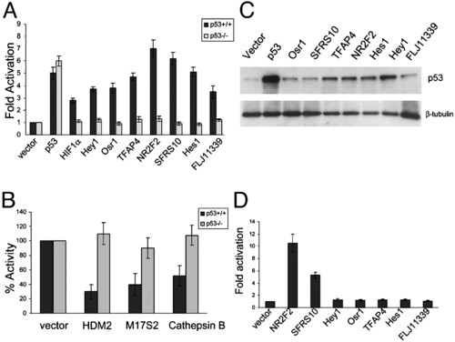
Novel p53 regulators activate or suppress p53 expression activity in mammalian cells. p53-positive regulators and control HIF1α (A) or negative regulators and control HDM2 (B) were transfected with FuGENE 6 into isogeneic HCT116 p53+/+ and p53-/- cells with the p53 reporter and a plasmid overexpressing β-galactosidase (β-gal). After 48 h incubation, cells were lysed, and luciferase activity was normalized based on β-gal activity. (C) Cell lysate from HCT116 cells transfected with p53 regulators were probed with antibodies against p53. (D) Reporter plasmid p53-1-luc was transfected into HCT116 p53-/- cells with p53 activators and β–gal plasmid. After incubating for 48 h, cells were lysed, and luciferase activity was normalized based on β-gal activity. |
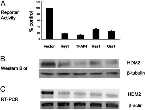
p53 regulators activate p53 by an HDM2-dependent mechanism. (A) A reporter plasmid containing the HDM2 promoter sequence was transfected into HCT116 p53-/- cells along with Hey1, Hes1, TFAP4, or Osr1 and β-gal plasmid. After incubating for 48 h, cells were lysed, and luciferase activity was detected and normalized based on β-gal activity. (B) Cell lysates from HCT116 p53-/- cells transfected with Hey1, Hes1, TFAP4,or Osr1 and immunoblotted with anti-HDM2 antibody. (C) mRNAs from HCT116 p53-/- cells transfected with Hey1, Hes1, TFAP4, or Osr1 were extracted, and RT-PCR was performed by using primers corresponding to HDM2 sequence. |
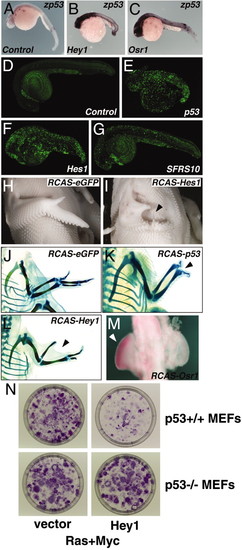
Phenotypic characterization of cDNAs in zebrafish, chicken, and MEFs. (A–M) Hey1, Hes1, Osr1. NR2F2, and SFRS10 induce p53-mediated apoptosis in vivo. (A–G) Zebrafish embryos were injected at one-cell stage with mRNAs encoding eGFP (control), p53, Hey1, Hes1, Osr1. NR2F2, or SFRS10 (B. C. F. and G), and analyzed at 30 h postfertilization (hpf) for p53 expression or at 40 hpf for cell death. Shown are representative images for different treatments and assays. (A–C) Detection of endogenous zebrafish p53 (zp53)by whole-mount in situ hybridization. Embryos injected with mRNAs encoding eGFP (A, control), Hey1 (B), or Osr1 (C) revealed strong induction of p53 transcripts, more widespread for Hey1 (B) than for Osr1 (C). Note the severe anterior and posterior defects induced by Hey1 overexpression (B). (D–G) Detection of apoptotic cells by acridine orange staining showed massive cell death in embryos injected with mRNAs encoding p53 (E) or Hes1 (F) whereas overexpression of SFRS10 (G) resulted in increased apoptosis in areas where normal programmed cell death takes place (D). Also note the severe anterior and posterior defects induced by p53 (E)or Hes1 (F) overexpression. All views are lateral, anterior to the left and dorsal to the top. (H–M) Chick embryos were injected in the prospective right wing field at HH14 (stage 14) with retroviruses (RCAS) encoding eGFP, p53, Hey1, Hes1, Osr1. NR2F2, or SFRS10 and analyzed at HH25 for cell death (M) or at HH35 for morphological wing alteration (H–L). Shown are representative images for different treatment and assays. (H and I) Injection of retroviruses encoding Hes1 (I) induced distal truncations in the right (injected) wing (arrowhead), compared with control-injected (H) or uninjected wings. (J–L) Cartilage staining showing typical skeletal patterns: embryos injected with RCAS-eGFP (J) showed a normal wing with three digits in the autopod, two well developed bones in the zeugopod, and one well developed bone in the stylopod. Embryo injected with RCAS-p53 (K) showed loss of digit III (arrowhead) and severe hypoplasia of digit II and IV. Embryos injected with RCAS-Hey1 (L) showed loss of digit II and defects in the zeugopod (arrowhead). (M) Staining with neutral red detected increased cell death in the distal mesenchyme of the wing bud injected with RCAS-Osr1 (arrowhead), compared with the contralateral, uninjected bud. (N) HEY1 suppresses ras/myc transformation in MEFs. MEFs from WT and p53 knockout mice were infected with retroviruses containing ras, myc, and Hey1 or vector. After 12 days of incubation, foci were stained with crystal violet and photographed. |