
- Title
-
Molecular structure and developmental expression of three muscle-type troponin T genes in zebrafish
- Authors
- Hsiao, C.-D., Tsai, W.-Y., Horng, L.-S., and Tsai, H.-J.
- Source
- Full text @ Dev. Dyn.

Tnnt temporal expression patterns in developing zebrafish embryos as detected by reverse transcriptase-polymerase chain reaction (RT-PCR). Total RNA was isolated from different developmental stages, and primers specific for sMyHC, Tnnt1, Tnnt2, Tnnt3a, and Tnnt3b were used for RT-PCR. β-actin was used as an internal control. Numbers in the right panel indicate the molecular mass of RT-PCR products. EXPRESSION / LABELING:
|
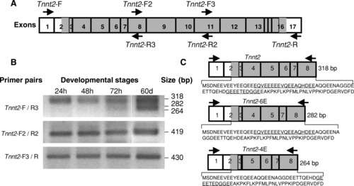
Reverse transcriptase-polymerase chain reaction (RT-PCR) analysis of Tnnt2 alternatively spliced variants during zebrafish development. A: Schematic representation of the positions of the primers used to perform RT-PCR. Empty boxes indicate untranslated regions; solid boxes show open reading frames. B: Age-dependent expression of alternatively spliced, Tnnt2 variants. Alternatively spliced variants were detected by RT-PCR using the primer pair combinations indicated. Two, alternatively spliced variants were detected by primer pair Tnnt2-F and Tnnt2-R3 in adult fish. C: Schematic illustration of the alternative splicing involved in the generation of Tnnt2 (unspliced form), Tnnt2-6E (excluding exon 6), and Tnnt2-4E (excluding exon 4). The alternatively spliced exons are underlined. |

Whole-mount in situ hybridization of Tnnt2 and other cardiac-specific genes during zebrafish cardiogenesis. Embryos were hybridized with Tnnt2 (A-K), cmlc2 (L), cmlc1 (M), vmhc (N), and cTnC (O) riboprobes, respectively. A-E: Lateral views, anterior of embryo to the left. F-I,K-O: Dorsal views, anterior of embryo to the top. J: Frontal view of embryo. Embryonic stages are indicated in each panel. h, hour; a, atrium; ht, heart tube; mp, myocardium precursor; v, ventricle. Scale bar = 100 μm in all pictures. EXPRESSION / LABELING:
|
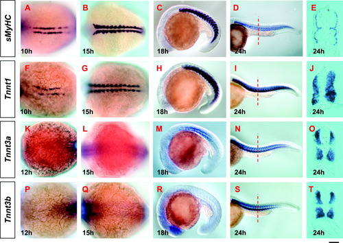
Whole-mount in situ hybridization of Tnnt1, Tnnt3a, Tnnt3b, and sMyHC during zebrafish early somitogenesis. Embryos were hybridized with sMyHC (A-E), Tnnt1 (F-J), Tnnt3a (K-O), and Tnnt3b (P-T) riboprobes, respectively. A,B,F,G,K,L,P,Q: Dorsal views. C,D,H,I,M,N,R,S: Lateral views. E,J,Q,T: Transverse sections at trunk level. The anterior is to the left in all whole-mount stained embryos. Dotted lines indicate the section level. Embryonic stages are indicated in each panel. h, hour. Scale bar = 200 μm in D,I,N,S, 140 μm in C,H,M,R, 100 μm in A,B,F,G,K,L,P,Q, 25 μm in E,J,O,T. |

Whole-mount in situ hybridization of Tnnt1, Tnnt3a, Tnnt3b, and sMyHC genes during zebrafish late somitogenesis. Embryos were hybridized with sMyHC (A-D), Tnnt1 (E-H), Tnnt3a (I-L), and Tnnt3b (M-P) riboprobes, respectively. C,G,K,O: Dorsal views. A,B,E,F,I,J,M,N: Lateral views. D,H,L,P: Ventral views. The anterior is to the left in all pictures. Embryonic stages are indicated in each panel. am, adductor mandibulae; fb, fin bud; hh, hyohyoideus; hm, horizontal myospetum; sh, sternohyoideus; sm, somites. Muscle nomenclature is based on Schilling and Kimmel ([1997]). Scale bar = 50 μm in B,F,J,N, 100 μm in A,C-E,G-I,K-M,O,P. |