
- Title
-
Retinoic acid signalling in the zebrafish embryo is necessary during pre-segmentation stages to pattern the anterior-posterior axis of the CNS and to induce a pectoral fin bud
- Authors
- Grandel, H., Lun, K., Rauch, G.J., Rhinn, M., Piotrowski, T., Houart, C., Sordino, P., Küchler, A.M., Schulte-Merker, S., Geisler, R., Holder, N., Wilson, S.W., and Brand, M.
- Source
- Full text @ Development
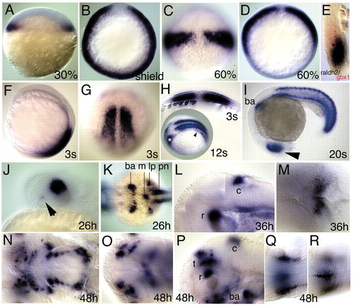
Whole-mount in situ hybridisations showing raldh2 expression in the zebrafish embryo and larva at (A) 30% epiboly, (B-E) gastrula, during (F-I) somitogenesis, and (J-R) larval stages. (A) Marginal view, animal pole is upwards. (B,D) Animal view, dorsal is upwards. (C,G) Dorsal view, animal/anterior is upwards. (E) Sagittal section along the animal vegetal axis. (F) Lateral view, anterior is upwards. (H) Cross section perpendicular to anteroposterior axis. (H inset) Dorsolateral view, anterior is to the left. (I,J,L,P) Lateral view, anterior is to the left. (K,M,O,Q,R) Dorsal view, anterior is to the left. (N) Ventral view, anterior is to the left. Note (A) the continuous expression in the blastoderm margin, (B) the exclusion from the shield and (C,D,E) the restriction to the involuting paraxial mesendoderm at 60% epiboly. (E) The sagittal section also reveals positioning of gbx1 (K. L. and M. B., unpublished) (Rhinn and Brand, 2001) in the neuroectoderm adjacent to raldh2. (F,G,H,I) Expression in the somites during somitogenesis stages. (H inset) Expression in the lateral plate mesoderm (arrowhead) extends into the prospective caudal part of the branchial arch primordium (asterisk) at 12s. (I,J) Dorsal expression in the retina (arrowhead) at 20s (I) and 26 hours (J) and weak ventral expression near the choroid fissure at 26 hours (arrowhead). (K) Expression in the caudal part of the branchial arch primordium (ba); in a mesenchymal domain in the midline below the notochord (m), in the anterior part of the pronephric ducts (pn) and in the posterior fin bud- and lateral plate-mesoderm (lp) at 26 hours (arrowhead). (L,M) Expression in the retina (r) and cerebellum (c) at 36 hours. (N-P) Expression at 48 hours. (N) In patches indicating the developing arches; (O) in four domains in the tectum and (P) in the retina (r), cerebellum (c), tectum (t) and branchial arches (ba). (Q,R) Expression surrounding the neural tube at the level of somites 3 to 4. EXPRESSION / LABELING:
|

Phenotype of nof homozygotes; wt: wild-type sibling. In A-I,K,M, anterior is to the left; in J,L,N proximal is to the left. (A,C) Lateral, (B,D) dorsal, (E-N) ventral views. (A-D) Larvae on day 5; ga, gill arches; pc, pericardial cavity; swb, swim bladder. (E,G) Branchial arch primordium of 36 hours embryos. (F) Presence and (H) absence of pectoral fin bud and AER marker dlx2 at 28 hours. (I,K) Cartilage pattern in heads and (J,L) fins of day 5 larvae. M, mandibular and H, hyoid arches; 3-7, gill arches; c, cleithrum; d, distal fin skeleton; g, proximal pectoral girdle. (M) Rescue of arch cartilages and (N) pectoral fins by treatment of nof homozygotes with 10–9 M retinoic acid at 30% epiboly until 16 hours. |

Anterior-posterior patterning defects in the CNS of nof mutants. In situ hybridisation as indicated of wild-type (A,C,E,G,I,K) and nof mutant (B,D,F,H,J,L) embryos at the 20s stage. (C,D,G,H) Curved red lines indicate the extent of tissue between different gene expression domains in wild type; arrowheads point to the borders of gene expression in wild type and nof mutants. EXPRESSION / LABELING:
|

Dorsal view of pectoral fins on day 5 in (A) a wild-type larva and (B) a nof homozygote after treatment with retinoic acid (10–9 M, 30% epiboly until 16 hours) and (C) after injection of wild-type raldh2 mRNA on day 6. (D) Injection of nof raldh2 mRNA does not provoke fin development in nof mutants. (E,F) Pectoral fins on d3 are prominent in wild type (E) but do not develop after injecting a raldh2-specific morpholino oligonucleotide (F). hoxb4a expression at 20s in wild-type (G) and (H) nof sibling embryos. (J,L) Similar hoxb4a expression levels as in nof homozygotes are detected upon injection of raldh2-specific morpholino into wild-type embryos (J) and upon injection of 500 pg mRNA derived from the nof-allele of raldh2 into nof homozygotes (L). (I,K) Increase of hoxb4a expression levels upon treatment of nof homozygotes (I) with retinoic acid (10–9 M; 30% until 20s) and (K) injection of 100 pg of wild-type raldh2 mRNA. EXPRESSION / LABELING:
|

(A-D) Dorsal view of the pectoral fin region on day 5 in (A) wild-type siblings and (B-D) nof mutant embryos. nof homozygotes were either (B) not treated or (C,D) treated with 10–9 M retinoic acid during the time windows indicated in the table above A-D. (E-H) Ventral view of the pectoral fin buds of (E) wild-type sibling, (F) nof and (G,H) BMS493-treated wild-type embryos. (E,H) Fin buds express dlx2, indicating AER activity in the apical ectoderm. (F,G) Fin bud regions do not express dlx2, indicating lack of AER activity in the ectoderm. (I,J) Dorsal view of the pectoral fin region of BMS493-treated wild-type embryos. Embryos were exposed to 10–6 M BMS493 at the different time windows indicated in the table above E-J. |

In situ hybridisations at the 20s stage to visualise the effects of treating wild-type embryos with 10–6 M BMS493 during different time windows on the hindbrain and the spinal cord. All panels are lateral views, anterior is to the left. Curved red lines indicate the lengths of hindbrain segments of untreated wild types in all panels. Black arrowheads indicate the true extend of hindbrain segments observed in untreated wild-type (A-C) and experimental embryos. (A,D,G) Expression of fgf8 and hoxb4a; (B,E,H) val and hoxb6b and (C,F,I) krx20 and hoxb5a. (D,E,F) Inhibition of RA signalling by BMS493 from 30% epiboly onwards expands the hindbrain between the fgf8 and hoxb4a domains, as well as val and krx20 domains; (D,E,F) hoxb4a, hoxb6b and the anterior part of the hoxb5a domain (red arrowhead) are strongly downregulated in the spinal cord. (G,H,I) BMS493 treatments that exclude pre-segmentation stages neither lead to expansion of the hindbrain nor to strong reduction of hoxb4a, hoxb6b and hoxb5a. |

In situ hybridisations at tail bud stage of (A,E,I) wild-type embryos and (B-D,F-H,J-L) embryos with compromised RA signalling. Dorsal views, anterior is to the top. Red lines refer to distances observed in wild-type embryos and are of the same length within each row of embryos. Homozygous nof, 5x10⁻⁶ M BMS493-treated or morpholino-injected embryos downregulate hoxb1a (B-D) and hoxb1b (F-H) expression compared with wild-type siblings (A,E). (I-L) Length of the prospective hindbrain territory between pax2.1 and val domains is longer in homozygous nof, 5x10⁻⁶ M BMS493-treated or morpholino-injected embryos (J-L) than in wild types (I) at the end of gastrulation. EXPRESSION / LABELING:
|

Unillustrated author statements |