
- Title
-
Removal of dystroglycan causes severe muscular dystrophy in zebrafish embryos
- Authors
- Parsons, M.J., Campos, I., Hirst, E.M.A., and Stemple, D.L.
- Source
- Full text @ Development

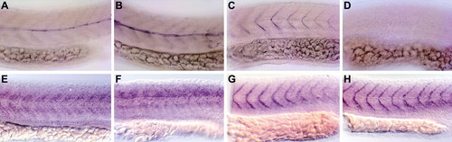
Whole-mount in situ hybridisation for dystroglycan. (A) 128-cell stage and (B) 1000-cell stage, indicating maternal expression. (C) Shield stage and (D,E) tailbud stage; posterior (D) and anterior (E) views, indicating expression in the chordamesoderm and developing brain. (F,G) Five-somite stage, posterior (F) and anterior (G) views. (H) Flat mount of a 12-somite embryo, showing widespread expression. (I) Sections through tailbud and hindbrain level of the 12-somite embryo in H. Expression in immature (upper section) but not mature (lower section) notochord is indicated (arrow). (J) Trunk of 24 hpf embryo. High expression in the hypochord is indicated (arrow). |
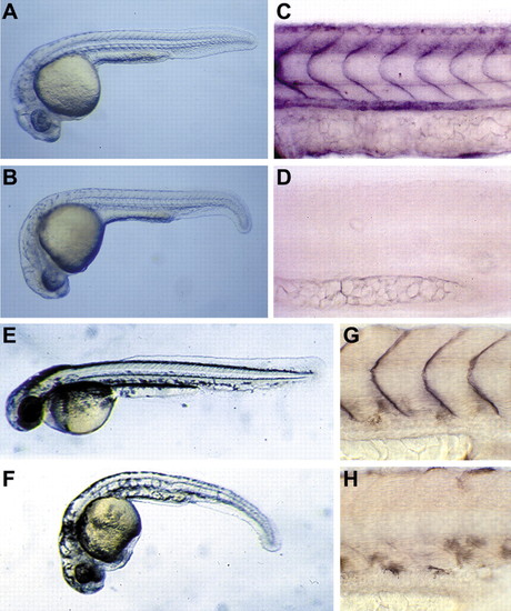
Removal of dystroglycan protein leads to abnormal development. Embryos injected with control MO at 24 hpf (A) and 48 hpf (E) and with a dystroglycan MO at 24 hpf (B) and 48 hpf (F). (C,D,G,H) Staining for dystroglycan in control (C,G) and dystroglycan morphant (D,H) embryos at 24 hpf (C,D) and 48 hpf (G,H). |
Molecular characterisation of the dystroglycan morphant phenotype. (A,C,E,G) Embryos injected with a control MO. (B,D,F,H) Dystroglycan morphants. (A,B) Staining for slow twitch myosin. Dystrophin is clearly localised to myosepta of control-MO-injected embryos (C) and is undetectable in dystroglycan morphants (D). Dystroglycan morphants do not show a decrease of either dystroglycan (E,F) or dystrophin (G,H) mRNA expression. EXPRESSION / LABELING:
|
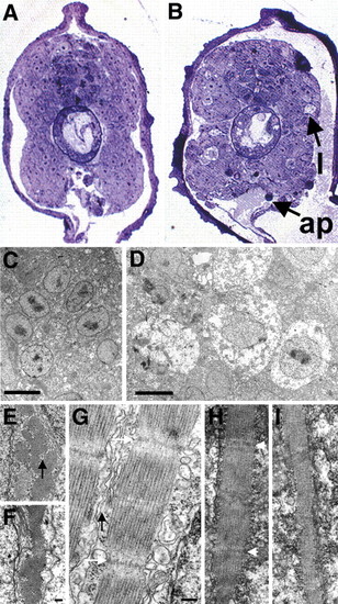
Histological analysis of dystroglycan morphants. Sections of control MO-injected (A,C,E,G) and dystroglycan morphant (B,D,F,H,I) embryos, all at 28 hpf. (A,B) Light micrographs of transverse sections. l, lesions; ap, apoptotic cells. (C,D) Electron micrographs of transverse sections through muscle. Lesions seen in B are confirmed as swollen, necrotic cells shown in D. (E-I) Subcellular structure of muscle cells at higher magnification. (E,F) Typical hexagonal array of actin and myosin in transverse sections. (G-I) Longitudinal sections. Z-discs are indicated, where detected, by white arrows. In the few identifiable sarcomeres in dystroglycan morphants, muscle fibre organisation is disrupted and sarcoplasmic reticulum is difficult to discern (H). The majority of muscle fibres in dystroglycan morphants possessed no sarcomeres (I). Sarcoplasmic reticulum in control embryos is indicated by black arrows. Scale bars: 5 μm in C,D; in F, 0.2 μm for E,F; in G, 0.2 μm for G-I. PHENOTYPE:
|

CNS and neuromuscular junction organisation is unaffected in dystroglycan morphants. Acetylated tubulin staining shows the pattern of axonal scaffolds in control MO-injected (A,B) and dystroglycan morphant (D,E) embryos. (A,D) Hindbrain views. (B,E) Anterior views. We could find no consistent morphological difference between control MO-injected (C) and dystroglycan morphant (F) after staining for AChRs. All embryos are at 48 hpf. PHENOTYPE:
|

Expression of laminin 1 and dystroglycan are mutually independent. Expression of laminin 1 in control MO-injected (A), dystroglycan morphant (B) and sly mutant (C) embryos. In the absence of dystroglycan, laminin 1 is still localised to the transverse myoseptum (B). Expression of dystroglycan in wild-type (D) and sly mutant (E) embryos. As indicated in C, sly mutants lack laminin 1, which does not affect dystroglycan localisation to the transverse myoseptum (E). EXPRESSION / LABELING:
|