
- Title
-
Bone patterning is altered in the regenerating zebrafish caudal fin after ectopic expression of sonic hedgehog and bmp2b or exposure to cyclopamine
- Authors
- Quint, E., Smith, A., Avaron, F., Laforest, L., Miles, J., Gaffield, W., and Akimenko, M.-A.
- Source
- Full text @ Proc. Natl. Acad. Sci. USA

(A) Schematic representation of the skeleton of a ray in the zebrafish fin. Dermal bone forms two segmented, concave and opposing hemirays. The diagram illustrates the position of amputations, relative to the bifurcation of the lepidotrichia; "low cut" and "high cut" are performed one to two segments proximal and four to six segments distal to the origin of bifurcation, respectively. (B) Sequence of shh expression patterns after a low cut, leading to ray bifurcations: (B1) 2 dpa, an unbifurcated ray expresses one shh domain in the distal blastema; (B2) by 4-5 dpa shh is expressed in two domains, preceding the formation of a bifurcation; (B3) two newly formed sister rays each exhibit one shh expression domain; (B4) the process repeats itself to form a second round of bifurcations (not to scale). (C) Ectopic expression of EGFP 24 h after injection of the pcmv5EGFP reporter construct in the tissue separating the branches of lepidotrichia (L) of two adjacent rays. Injections were performed at 2 dpa (high cut shown by the dotted line). (D-I) Normal bifurcating (D, E) and N-shh (F, G) or bmp2b (H, L) injected fused rays stained with Alcian blue. Constructs were injected in the region indicated by white arrows (F, H). (D, F, H) Whole-mount control and fused rays; proximal part of the fin is to the left. (E, G, I) Cryosections (16 μm) in the region of normal bifurcation (E) or fusion (G, I). Arrows in G and I indicate the region of ectopic bone matrix deposition. No ectopic bone deposition occurs in the mesenchyme interior of the lepidotrichia (L) as marked with asterisks. (Bar = 50 μm.) |

Transverse sections of regenerating fins showing the normal expression patterns of shh (A), ptc1 (B) in two adjacent rays; bmp2b (C) in one ray at 4 dpa; ptc1 expression 2 days after injection of N-shh (D) or bmp2b (E) constructs. Note the ectopic expression of ptc1 in mesenchyme and basal epithelial cells separating the branching rays (indicated by the brackets) after ectopic expression of N-shh (D) but not bmp2b (E). Arrows in A-C indicate shh, ptc1, and bmp2b expression in basal epithelial cells. Arrowheads in B and C indicate ptc1 and bmp2b expression in scleroblasts. (Bar = 50 μm.) |
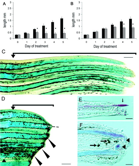
(A, B) Growth curves of fins treated with 10 μM (A) or 5 μM (B) solanidine (black bars) or cyclopamine (diagonal hatched bars). The ordinate indicates the length of the fins in millimeters and the abscissa the day of treatment, where day 0 is the day fish were first put into alkaloid solution. At 10 μM, fin outgrowth is significantly inhibited by day 2 of treatment (asterisks, P < 0.05) and by day 4 of treatment at 5 μM (asterisks, P < 0.05). P values were derived by using the Student's t test, error bars = SEM. (C-F) Effects of cyclopamine on caudal fin regeneration, proximal part of the fin is to the left; (C, D) whole mount fins stained with Alcian blue. Exposure to 5 μM cyclopamine (D) for 5 days results in the inhibition of fin outgrowth compared with 5 μM solanidine (C). Melanocytes accumulate in the distal stumps of the rays (large arrowheads) and the blastema is clearly absent. Small arrowhead in D indicates the level of fin amputation (low cut). The dotted line indicates the plane of sections shown in E and F. Cryosections (16 μm) along the longitudinal axis of fins treated with 10 μM cyclopamine (F) or solanidine (E) and stained with Alcian blue and Sirius red. Note pigment accumulation and extra bone deposition in the distal-most portion of the section (arrows) and the lack of actinotrichia as shown in E (arrow). [Bar (C, D) = 100 μm; (E, F) = 25 μm.] |

Gene expression in regenerating caudal fins treated with 5 μM solanidine (A, C, E) or cyclopamine (B, D, F). After 1 day of cyclopamine, ptc1 expression, normally expressed in a single domain (A), is absent (B) and shh, normally expressed in two domains (C, E, arrows) is present as a single and expanded domain along the proximal-distal axis (D). By day 3 of treatment, cyclopamine-treated fins show only one weak and diffuse domain of shh expression (F, arrows) in the region of pigment accumulation and lepidotrichia termination (square brackets). (Bar = 150 μm.) |

BrdUrd-incorporation in fins treated with cyclopamine or solanidine. After 1 day of treatment (initiated at 2 dpa) with either solanidine (A) or cyclopamine (B), proliferating cells were identified in the epidermis (solid arrows) and mesenchyme (arrowheads) in the region of new bone matrix secretion. By day 3, BrdUrd-labeled cells were still found in the epidermal layers of both control and cyclopamine-treated fins (C, D, solid arrows) and in the mesenchyme of solanidine-treated fins (C, arrowheads), but not in the mesenchyme of cyclopamine-treated fins (D). Dotted lines mark the level of amputation. (Bar = 50 μm.) |