
- Title
-
zic gene expression marks anteroposterior pattern in the presumptive neurectoderm of the zebrafish gastrula
- Authors
- Grinblat, Y. and Sive, H.
- Source
- Full text @ Dev. Dyn.

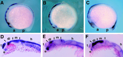
zic gene expression is patterned in the neurectoderm during gastrulation. Staged embryos were stained for opl/zic1, zic2, zic3 (purple), and otx2 RNA (orange in G-I) by using whole-mount in situ hybridization. A-C: Early gastrula (shield) stage embryos, dorsal view, anterior at the top. A: opl/zic1 is not expressed. B: zic2 is expressed in the blastoderm margin, the presumptive mesendoderm, only. C: zic3 expression in the posterior portion of the dorsal ectoderm and in the blastoderm margin. D-F: Mid-gastrula (80% epiboly) stage embryos, dorsal view, anterior at the top. D: opl/zic1 expression restricted to an anterior domain of the neural plate. E: zic2 expression domain includes the anterior opl domain and extends more posteriorly. F: zic3 is expressed in the posterior portion of the neural plate and at the edge of the neural plate anteriorly. G-I: Late gastrula (90% epiboly) stage embryos, dorsal view, anterior at the top. G,H: Expression of opl (G) and zic2 (H), shown in purple, overlap the otx2 (orange) expression domain in the presumptive forebrain. I: anterior portion of zic3 expression overlaps the posterior portion of the otx2 domain. Dots indicate outline of the blastoderm margin. White arrowheads point to the posterior extent of otx2 staining, which corresponds to the future posterior midbrain. EXPRESSION / LABELING:
|
Patterned zic gene expression in the dorsal brain during somitogenesis. Staged embryos were stained for opl/zic1, zic2, or zic3 RNA (purple) by using whole-mount in situ hybridization. A-C: Mid-somitogenesis (15 hpf) embryos, lateral view, anterior to the left, dorsal at the top. A: opl/zic1 is expressed in the dorsal brain and in the dorsal half of each somite (Rohr et al., [1999]). B: zic2 is expressed in the dorsal brain, the forming optic stalk, and weakly in the developing tailbud. C: zic3 is expressed in the dorsal brain and strongly in the tailbud; also note transient expression in the ventral diencephalon. D-I: Late somitogenesis (prim-5) embryos, side view, anterior to the left, dorsal at the top. D: opl/zic1 is expressed throughout the dorsal brain and in the optic stalk. E: zic2 expression is restricted to the dorsal diencephalon, dorsal portion of the cerebellum, and the optic stalk (confirmed in sections, data not shown). F: zic3 is expressed in the dorsal diencephalon, tectum, and the optic stalk. a, anterior; p, posterior; t, telencephalon; di, diencephalon; m, midbrain; h, hindbrain; asterisks indicate optic stalk. EXPRESSION / LABELING:
|

Early pattern in the ectoderm, marked by zic3 expression, is regulated by bone morphogenetic protein (BMP) signaling. Embryos stained by whole-mount in situ hybridization (ISH) for expression of zic3 (orange in A, purple in C-F) and bmp2b/swr (purple in A). All embryos are shown in animal pole view, dorsal to the right. A: zic3 (orange) and bmp2b/swr (purple) are expressed in complementary domains in wild-type embryos. B: Diagram of a specification map, derived from in vitro explant culture assays, of the early gastrula ectoderm (Grinblat et al., [1998]). V, ventral non-neural ectoderm; A, anterior forebrain; P, posterior forebrain. C: Embryo derived from a din/+x din/+ cross, showing a wild-type pattern of zic3 expression. D: Sibling embryo from the same cross, showing a strong reduction in dorsal zic3 expression. Dorsal marginal staining remained unaffected (not shown). E: Embryo derived from a snh/+ x snh/+ cross, showing a wild-type pattern of zic3 expression. F: Sibling embryo from the same cross, showing up-regulation of zic3 expression in ventral posterior ectoderm (white asterisk). Note that the animal pole is still devoid of zic3 expression. d, dorsal; white asterisk marks ventral side. |

Unillustrated author statements |