
- Title
-
Zebrafish genes rx1 and rx2 help define the region of forebrain that gives rise to retina
- Authors
- Chuang, J.-C. and Raymond, P.A.
- Source
- Full text @ Dev. Biol.

Expression of rx genes during neurulation in wild type and cyclops embryos. (A, B) Normal expression pattern of rx in the rostral neural plate (A) in relation to emx1 expression or (B) by itself. (C) Expression of rx3 in cyclopsb16 embryos at the neural plate stage. Note the absence of the caudal indentation at the midline [compare arrow in (C) with arrows in (A) and (B)]. (D-G) Transverse sections of whole-mount, double in situ preparations at the level of (D) the retinal field or (E-G) the optic primordia. Note the relationship between retinal precursors (marked by rx, in red) and telencephalic precursors (marked by emx1 or BF-1, in purple). The unlabeled cells in the ventral neural keel are ventral diencephalic precursors. (H-K) Comparison of rx1 expression in wild type (H, J) and cyclops embryos (I, K). The arrows indicate the suppression of rx1 expression at the caudal midline. The arrowheads indicate the region in which rx1 expression differs between wild type and cyclops embryos. Note that rx1 is not expressed in the rostral hypothalamus in wild type embryos, as reflected by an indentation at the midline on the rostral side (arrowhead). In cyclops embryos (I, K), the rostral hypothalamus is missing, and the two eyes are fused in the rostral midline (arrowhead). Rostral is up in (A-C) and (H-K). op, optic primordium; t, telencephalon; vd, ventral diencephalon. Scale bars: 100 μm. EXPRESSION / LABELING:
|

Eye phenotypes and axis duplication in embryos injected with rx1 or rx2 RNA. (A-J) Comparison of wild type (A, C, E, G) and rx1- or rx2-injected embryos (B, D, F, H–J) at 60 h postfertilization (hpf), in a ventral view (A, B, E, F, J), a lateral view (C, D), or a frontal view (G-I). Arrows indicate the medial expansion of retinal tissue toward forebrain territories. (K) Partial axis duplication (arrows) caudal to the midbrain. (L) Transverse section showing a third eye in the ventral midline (arrow) and a partially duplicated forebrain (arrowheads). (K) and (L) were hybridized with a pax6.1 RNA probe and viewed in a whole-mount (K, 60 hpf) or in a cryosection (L, 26 hpf). cf, choroid fissure; d, diencephalon; f, forebrain; h, hindbrain; L, lens; m, midbrain; sc, spinal cord; t, telencephalon; vd, ventral diencephalon. Scale bars: 100 μm. |

Morphology of eye and brain of living embryos between 24 and 37 hpf. (A-C) Comparison of control (uninjected) and injected embryos at 24 hpf (dorsal view). In the control embryo (A), the two eyes are separated by the neural tube at the midline. In injected embryos (B, C) the eyes have expanded across the midline (arrowhead), resulting in deletion of forebrain (telencephalon). In (B), the eyes have fused with the diencephalon (arrows) proximal and caudal to the optic stalks. In (C), the eyes have fused at the midline, but also remain attached to the diencephalon through the optic stalks, in the region of the hypothalamus. The ventricular space in the midbrain is enlarged in this embryo. (D-F) Comparison of control, injected, and cyclops embryos at 37 hpf (frontal view). In the injected embryo (E), the expanded eyes have obliterated the telencephalon, but the hypothalamus is present. In contrast, the cyclops embryo (F) lacks a hypothalamus but retains the telencephalon. Embryos were injected with 300 pg rx2ΔRx (B, C) or 1 ng rx2ΔC′ (E) RNA. d, diencephalon; e, eye; h, hypothalamus; m, mesencephalon; os, optic stalks; t, telencephalon. Scale bars: 100 μM. |

Selective loss of forebrain structures in embryos following rx2 overexpression. (A-D) Comparison of shh and rx3 expression in control and injected embryos at 24 hpf (A, B; lateral view) or 60 hpf (C, D; transverse section). (C) rx3 is expressed in the inner nuclear layer of the neural retina and in the hypothalamus. (E-J) Comparison of crx, BF-1, and emx1 expression in control and injected embryos at 36 hpf (E-H) or 24 hpf (I, J). (E) and (F) are lateral views; (G-J) are dorsal views. Note the absence of crx expression in the epiphysis in (F), the strong suppression of BF-1 in the telencephalon in (H), and the absence of emx1 expression in (J). Arrows in (E) and (F) indicate strong expression of crx in the earliest differentiating retina, located at the ventronasal edge. (K, L) Anti-acetylated α-tubulin (α-tub) staining (dorsal view, 24 hpf). Black arrows show the major axonal tracts in the forebrain and white arrowheads show the axonal tracts in the midbrain region. Embryos were injected with 1 ng rx2ΔC′ (B, D, J, L), or 300 pg rx2ΔRx (F, H) RNA. d, diencephalon; ep, epiphysis; h, hypothalamus; INL, inner nuclear layer; t, telencephalon; tec, optic tectum; V, ventricle; vd, ventral diencephalon; vnr, ventral neural retina. Scale bars: 100 μm. |
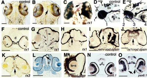
Abnormalities of eye development following rx2 overexpression. (A, B) Comparison of eye morphology in control and rx2-injected embryos (PTU-treated, dorsal view) at 88 hpf. The arrow in (B) indicates medial expansion of the right eye. (C) Ectopic expression of rx1 (arrowheads) in the dorsal forebrain. (D, E) Comparison of control and rx2-injected embryos (lateral view). Lines in (D) and (E) represent the estimated section planes shown in (F, N): 1; (H): 2; (G, I–L, O): 3; and (M): 4. (F) In control embryos at 74 hpf, rx1 is expressed in the outer nuclear layer and circumferential germinal zone (arrowheads) of the neural retina. (G) Ectopic cone photoreceptors (expressing rx1) form rosettes within neural retina (arrowheads) and are found at the boundary between the fused eyes (arrow). (H) The entire rostral end of this embryo (arrow) consists of retinal tissue. (I) Ectopic RPE and photoreceptors (arrow) are found in forebrain, and ectopic RPE is in neural retina (arrowhead). (J) Ectopic photoreceptors (arrow) are in forebrain. (K) No forebrain tissue is found between the two eyes (arrow). In the differentiated retina, rx3 is expressed in the inner nuclear layer (Chuang et al., 1999). (L) Replacement of telencephalic tissue by ectopic RPE (arrow; methylene blue staining). (M) Ectopic RPE is surrounding the lens. (N–O) Comparison of pax6.1 expression patterns between control and injected embryos. The arrows in (O) show the lack of RPE and expansion of neural retina ventrally. Embryos were injected with 300 pg rx2ΔRx (B, E), 600 pg myc-rx2 (C, G–J, L, M), or 50 pg rx2ΔN′ (K, O) RNA, and examined at 88 hpf (A, B), 78 hpf (C, G, J), 60 hpf (D, E, N, O), 74 hpf (F, K), 96 hpf (H, I, M), and 46 hpf (L). d, diencephalon; mhb, midbrain hindbrain boundary; ONL, outer nuclear layer; ov, otic vesicle; t, telencephalon; V, ventricle. Scale bars: 100 μm. EXPRESSION / LABELING:
|
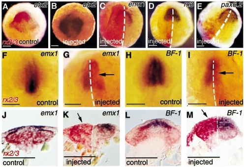
Gene expression patterns at neural plate and neural keel stages. Red staining represents endogenous rx2 and/or rx3 (A, F, H, J, L) and ectopic rx2 (B-E, G, I, K, M) expression. Purple staining represents the expression of otx2 (A, B); emx1 (C, F, G, J, K); rx3 (D); pax6.2 (E); or BF-1 (H, I, L, M). Note suppression of emx1 (G, K, arrows) or BF-1 (I, M, arrows) by ectopic rx2. Embryos in (B-E, G, I, K, M) were injected with 1 ng rx2ΔC′ RNA. Dorsal view of embryos at 9-10.5 hpf (A-E), or 11-13 hpf (F-M). (J-M) Transverse sections of the whole-mounted embryos are shown in the above panels, respectively. Rostral is up in (A-I). Scale bars: 100 μm. |

Ectopic expression of eye-determination genes, cell death, and proliferation in rx2-injected embryos. (A-C) Normal expression patterns. (E–G) Ectopic expression of eye-determination genes (arrows). Note in (C) and (G), purple/brown staining shows the endogenous rx2 expression (detected by a probe transcribed from 3′ coding and 3′ UTR sequences that were not present in the injection construct, rx2DC9). In (G), red staining shows the exogenous rx2 (detected by a probe transcribed from rx2ΔC′). (D, H) Dorsal view of living embryos stained with acridine orange. (H) Note more cell death on the left side of the injected embryo. (I-K′) Transverse sections of embryos showing expression of rx1 (I, J, K) and Cy-3 (red fluorescence) staining of mitotic cells and DAPI nuclear staining (I′, J′, K′) of the same sections, respectively. The arrows in (J-K′) indicate ectopic expression of rx1 in forebrain territory in injected embryos. Embryos were injected with 300 pg β-galactosidase (D), 1 ng rx2ΔC′ (E, G), 300 pg rx2ΔRx (H, J-K′), or 600 pg myc-rx2 (F) RNA, and examined at 16 hpf (A-B, E-F), 15 hpf (C, G), or 19 hpf (D, H, I-K′). Rostral is up in (A-H). α-PH3, anti-phosphohistone H3 antibody. Scale bars: 100 μm. EXPRESSION / LABELING:
|
Reprinted from Developmental Biology, 231, Chuang, J.-C. and Raymond, P.A., Zebrafish genes rx1 and rx2 help define the region of forebrain that gives rise to retina, 13-30, Copyright (2001) with permission from Elsevier. Full text @ Dev. Biol.