
- Title
-
The vascular anatomy of the developing zebrafish: an atlas of embryonic and early larval development
- Authors
- Isogai, S., Horiguchi, M., and Weinstein, B.M.
- Source
- Full text @ Dev. Biol.
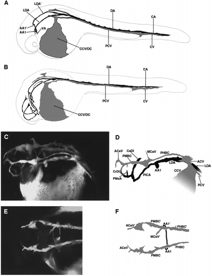
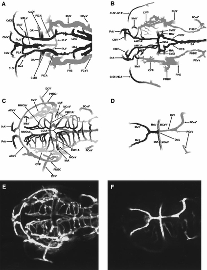
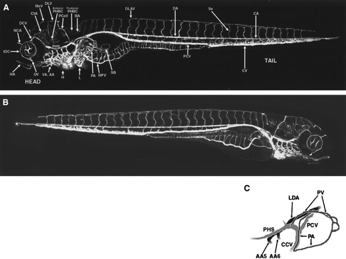
Circulation in the developing zebrafish at 24–28 h postfertilization. (A) Diagram of active vessels in the zebrafish embryo just after the initiation of circulation, at approximately 24 h postfertilization (hpf). A single circulatory loop is present passing obligatorily through the trunk tail. (B) Diagram of active vessels in the zebrafish embryo a short time later, at approximately 26 hpf. A second cranial circulatory loop is now present. The stages in (A) and (B) are depicted using drawings because the weak circulation at these stages makes acquisition of attractive angiographic images difficult. These patterns are, however, representative of those seen in angiograms. (C) Angiogram of the head of a developing zebrafish at approximately 28 hpf. Dorsal–lateral view. (D) Diagram of vessels in (C). Only the vessels on the left side are shown, for simplicity. The major, primary arterial and venous vessels of the head are visible. (E) Angiogram of the head of a developing zebrafish at approximately 28 hpf. Dorsal view. Only the major primary venous vessels (PMBC, PHBC) are visible. The primary arterial vessels (LDA, PICA) are present but are directly underneath and obscured by the PMBC and PHBC in this view. (F) Diagram of vessels in (E). All panels are oriented with rostral to the left, and all lateral views are from the left side. A glossary of the names corresponding to all labeled vessels is provided in Table 1. |
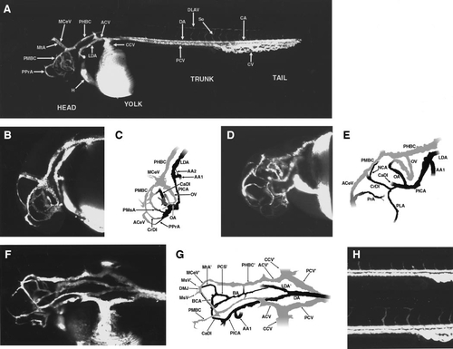
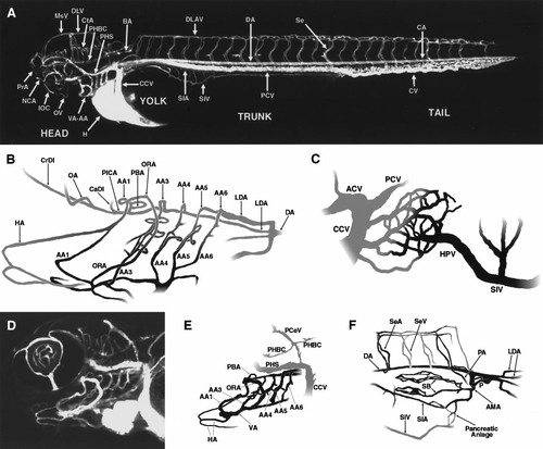
Circulation in the developing zebrafish at 1.2–1.5 days postfertilization (dpf). (A) Angiogram of a developing zebrafish at approximately 1.5 dpf, compiled from two separate reconstructions pasted together. Lateral view. (B) Angiogram of the head of a developing zebrafish at approximately 1.3 dpf. Lateral view. (C) Diagram of vessels in (B). Only the vessels on the left side are shown. (D) Angiogram of the head of a developing zebrafish at approximately 1.5 dpf. Ventral–rostral–lateral view. (E) Diagram of vessels in (D). Only the vessels on the left side are shown. (F) Angiogram of the head and cranial trunk of a developing zebrafish at approximately 1.5 dpf. Dorsal–lateral view. (G) Diagram of vessels in (F). (H) Angiogram of the caudal trunks of developing zebrafish at approximately 1.2–1.5 dpf. Lateral views. The emergence of the intersegmental (intersomitic) vessels of the trunk is shown. The upper angiogram shows a somewhat earlier stage than the lower angiogram. All lateral views are from the left side. A glossary of the names corresponding to all labeled vessels is provided in Table 1. |
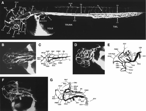
Circulation in the developing zebrafish at approximately 2 days postfertilization (dpf). (A) Angiogram of a developing zebrafish at approximately 2 dpf, compiled from three separate reconstructions pasted together. Lateral view, labeled. (B) Angiogram of the head of a developing zebrafish at approximately 1.8 dpf. Dorsal view. (C) Diagram of vessels in (B). Vascular connections are apparent between the BA and both PHBC. (D) Angiogram of the head of a developing zebrafish at approximately 2.0 dpf. Ventral–rostral–lateral view. (E) Diagram of vessels in (D). Only the vessels on the left side are shown, for clarity. Note that the PPrA has now disconnected from the CrDI and the PMsA has disconnected from the PMBC. (F) Angiogram of the head of a developing zebrafish at approximately 2.0 dpf. Dorsal–lateral view. The vessels shown are mostly those on the left side, for clarity. The major arterial and venous pathways of the head at this stage are apparent. (G) Diagram of vessels in (F). All panels are oriented with rostral to the left, and all lateral views are from the left side. A glossary of the names corresponding to all labeled vessels is provided in Table 1. |
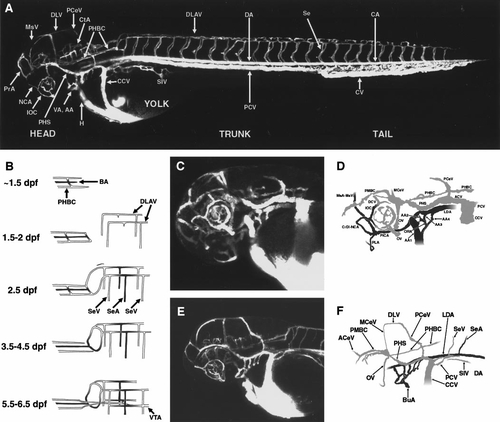
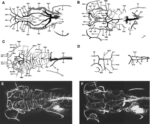
Circulation in the developing zebrafish at approximately 2.5 days postfertilization (dpf). (A) Angiogram of a developing zebrafish at approximately 2.5 dpf, compiled from three separate reconstructions pasted together. Lateral view, labeled. (B) Schematic diagram showing vessels of the caudal head and cranial trunk, how these vessels become connected, and how this changes over the course of several days. (C) Angiogram of the head of a developing zebrafish at approximately 2.5 dpf. Ventral–lateral view. (D) Diagram of vessels in (C). Only the vessels on the left side are shown, for clarity. AA1–AA4 have formed, and the PHS is now on line. (E) Angiogram of the head of a developing zebrafish at approximately 2.5 dpf. Lateral view. (F) Diagram of vessels in (E). Only the vessels on the left side are shown, for clarity. All six aortic arches are now carrying blood flow. All panels are oriented with rostral to the left, and all lateral views are from the left side. A glossary of the names corresponding to all labeled vessels is provided in Table 1. |
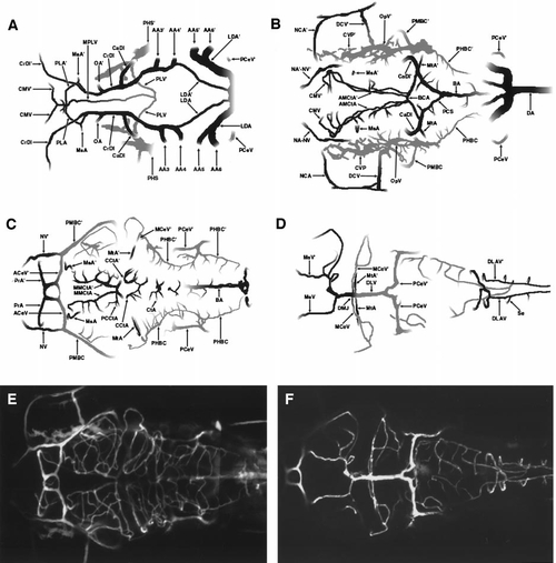
Multilayer composite diagram of circulation in the developing zebrafish head at approximately 2.5 days postfertilization (dpf). A 2.5 dpf dorsal–view angiographic image stack was divided into 4 substacks and each substack was separately reconstructed. Drawings were prepared detailing the vascular patterns in each of these reconstructions. (A) Bottom layer diagram, showing ventral cranial vessels (beginning approximately just above the future pharynx). (B) Lower middle layer diagram. (C) Upper middle layer diagram. (D) Top layer diagram, showing the most dorsal cranial vessels. (E) Angiogram corresponding to the diagram in panel (C). (F) Angiogram corresponding to the diagram in panel (D). All panels are oriented with rostral to the left and left down. A glossary of the names corresponding to all labeled vessels is provided in Table 1. |
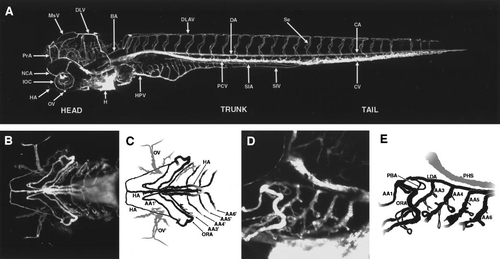
Circulation in the developing zebrafish at approximately 3–3.5 days postfertilization (dpf). (A) Angiogram of a developing zebrafish at approximately 3.5 dpf, compiled from four separate reconstructions pasted together. Lateral view, labeled. (B) Schematic diagram illustrating how the aortic arches feed into the ventral cranial arterial circulation, prepared from a Berlin-blue dye-injected 3.5 dpf specimen. Note that the caudal two aortic arches (AA5 and AA6) empty into a separate branch of the lateral dorsal aorta. (C) Diagram showing the renal vascular plexus and associated vessels that feed and drain the liver, at 3.5 dpf. (D) Angiogram of the head of a developing zebrafish at approximately 3 dpf. Ventral–lateral view. (E) Diagram of vessels in (D). Only the aortic arches, associated arterial vessels, and a few of the major venous vessels seen in (C) are depicted in the diagram. Note the appearance of the HA, and elongated VA, and vascular sprouts appearing on AA3–AA6. (F) Diagram showing the anterior mesenteric artery and its branches in the ventral cranial trunk at 3 dpf, prepared from a Berlin-blue dye-injected 3.5 dpf specimen. All panels are oriented with rostral to the left, and all lateral views are from the left side except for (F), which is a right-side view with rostral to the right. A glossary of the names corresponding to all labeled vessels is provided in Table 1. |
Multilayer composite diagram of circulation in the developing zebrafish head and cranial trunk at approximately 3.5 days postfertilization (dpf). A pair of 3.5 dpf dorsal-view angiographic image stacks were divided into 4 paired substacks, and each of these was separately reconstructed. Drawings were prepared detailing the vascular patterns in each of the four reconstructions. (A) Bottom layer diagram, showing ventral cranial vessels (beginning approximately just above the future pharynx). (B) Lower middle layer diagram. (C) Upper middle layer diagram. (D) Top layer diagram, showing the most dorsal cranial vessels. (E) Angiogram corresponding to the diagram in panel (C). (F) Angiogram corresponding to the diagram in panel (B). All panels are oriented with rostral to the left and left down. A glossary of the names corresponding to all labeled vessels is provided in Table 1. |
Circulation in the developing zebrafish at approximately 4 days postfertilization (dpf). (A) Angiogram of a developing zebrafish at approximately 4 dpf, compiled from five separate reconstructions pasted together. Lateral view, labeled. (B) Angiogram of the ventral head of a developing zebrafish at approximately 4 dpf. (C) Diagram of vessels in (B). In this ventral view only the aortic arches and adjacent vessels are visible. All extant aortic arches are shown, as well as the hypobranchial vessels. (D) Higher magnification (20x) angiogram of the ventral head of a developing zebrafish at approximately 4 dpf. Lateral view, showing primarily aortic arches. (E) Diagram of vessels in (D). Note the vascular loops associated with AA3–AA6. Separation of these AA into parallel afferent and efferent derivatives (ABA and ABF) is in progress but not well visualized in this particular orientation of the embryo. In all panels rostral is to the left, and all lateral views are from the left side. A glossary of the names corresponding to all labeled vessels is provided in Table 1. |
Circulation in the developing zebrafish at approximately 4.5 days postfertilization (dpf). (A) Angiogram of a developing zebrafish at approximately 4.5 dpf, compiled from five separate reconstructions pasted together. Left-side lateral view, labeled. (B) Angiogram of a developing zebrafish at approximately 4.5 dpf, compiled from five separate reconstructions pasted together. Right-side lateral view, unlabeled. (C) Diagram illustrating the pectoral artery (PA) and pectoral vein (PV), and the source (from the DA) and drainage (into the ACV) of these vessels. This diagram was prepared from the angiographic stack used to generate panel (A). All panels are oriented with rostral to the left, and all lateral views are from the left side except for (B), which is a right-side view with rostral to the right. A glossary of the names corresponding to all labeled vessels is provided in Table 1. |
Multilayer composite diagram of circulation in the developing zebrafish head and cranial trunk at approximately 4.5 days postfertilization (dpf). A pair of 4.5 dpf dorsal-view angiographic image stacks were divided into 4 paired substacks, and each of these was separately reconstructed. Drawings were prepared detailing the vascular patterns in each of the four reconstructions. For clarity, only major arterial and venous vessels and short connecting portions of smaller caliber vessels are shown in each diagram. (A) Bottom layer diagram, showing ventral cranial vessels (beginning approximately just above the future pharynx). (B) Lower middle layer diagram. (C) Upper middle layer diagram. (D) Top layer diagram, showing the most dorsal cranial vessels. (E) Angiogram corresponding to the diagram in panel (C). (F) Angiogram corresponding to the diagram in panel (D). All panels are oriented with rostral to the left and left down. A glossary of the names corresponding to all labeled vessels is provided in Table 1. |
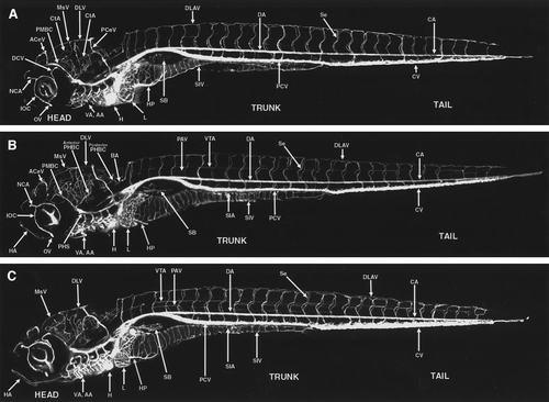
Circulation in the developing zebrafish at 5.5–7.5 days postfertilization (dpf). (A) Angiogram of a developing zebrafish at approximately 5.5 dpf, compiled from five separate reconstructions pasted together. (B) Angiogram of a developing zebrafish at approximately 6.5 dpf, compiled from five separate reconstructions pasted together. (C) Angiogram of a developing zebrafish at approximately 7.5 dpf, compiled from six separate reconstructions pasted together. All panels are oriented with rostral to the left and ventral down, and particular vessels have been labeled in each panel. A glossary of the names corresponding to all labeled vessels is provided in Table 1. |
Reprinted from Developmental Biology, 230(2), Isogai, S., Horiguchi, M., and Weinstein, B.M., The vascular anatomy of the developing zebrafish: an atlas of embryonic and early larval development, 278-301, Copyright (2001) with permission from Elsevier. Full text @ Dev. Biol.