
- Title
-
The zebrafish floating head mutant demonstrates podocytes play an important role in directing glomerular differentiation
- Authors
- Majumdar, A. and Drummond, I.A.
- Source
- Full text @ Dev. Biol.
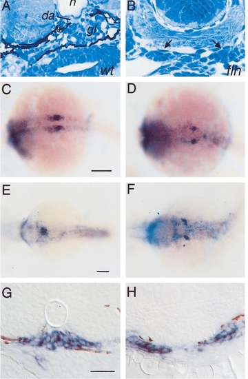
Glomerular development is altered in flh. Transverse histological sections (A, B). In wildtype zebrafish embryos at 2 days postfertilization (dpf) (A) a midline glomerulus (gl) is seen underneath the notochord (n) and dorsal aorta (da). A midline glomerulus is not seen in flh (B) at 2 dpf. Instead, clusters of cells are found in ectopic locations in flh (arrows in B). Whole-mount in situ hybridization with the wt1 probe (C-F). wt1 is expressed in nephric primordia in wildtype embryos at 20 h postfertilization (hpf) (C) and later at 48 hpf (E) in differentiated podocytes after pronephric primordia fuse in the midline. wt1 is expressed in flh nephric primordia at 20 hpf (D), but wt1-expressing cells remain in ectopic lateral positions at 48 hpf (F). Transverse sections of wt1 hybridized 36-hpf embryos (G, H). wt1-expressing cells are found at the midline in wildtype siblings (G) and in laterally positioned cells in flh (H). Some brown pigment granules are seen in these sections. Scale bars: (C, D) 200 µm; (E, F) 250 µm; (G, H) 50 µm. EXPRESSION / LABELING:
|

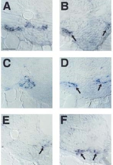
Glomerular defects in sonic you and you-too mutants correlate with failed sclerotome development. Transverse section of a 48-hpf syutbx392/t4 embryo (A) reveals the absence of a midline glomerulus and in its place are two lateral cell clusters (arrows). In situ hybridization of syutbx392/t4 embryos with a wt1 probe (B) identifies the lateral cell clusters as podocytes. Similar histological section of a 36-hpf you-too embryo (C) shows a failed convergence of podocytes. In situ hybridization with gli2 probe (D) shows expression in the neural tube and fin bud (dark arrows) but no expression in the pronephric primordia (light arrow). Transverse histological section through a wildtype 3-week-old zebrafish (E) shows the glomerulus (gl) is surrounded by bone and cartilaginous tissue (double arrows; da, dorsal aorta). twist-positive cells are found in the wildtype ventromedial somite at 18 hpf (F). twist-positive cells are absent in flh mutants (G). Pax9-expressing cells are found between neural tube and somite tissue in 24-hpf wildtype embryos (H). Pax9-expressing cells are reduced in flh (I). Scale bar equals 50 µm. EXPRESSION / LABELING:
PHENOTYPE:
|
Glomeruli differentiate at ectopic locations in flh. Wildtype (A, C, E) and flh (B, D, F) embryos hybridized with in situ probes: VEGF (A, B) at 40 hpf, flk-1 (C, D) at 40 hpf, and tie-1 (E, F) at 48 hpf. Differentiated podocytes express VEGF in wildtype embryos (A). VEGF-positive cells are found in ectopic lateral locations in flh (arrows in B). flk-1 labels glomerular capillary-forming endothelial cells in wildtype (C). flk-1-positive cells surround laterally positioned nephric primordia in flh (arrows in D). flk-1-positive cells are found in between nephric primordia and cardinal veins. tie-1 labels endothelia from cardinal veins (arrow in E) but not dorsal aorta in wildtype embryos at this time. In flh, tie-1-positive cells are associated with nephric primordia (arrows in F). Scale bar equals 50 µm. |

flh embryos make functional glomeruli as assayed by filtration of rhodamine dextran dye. Rhodamine dextran dye (red) has been injected into the sinus venosus, filtered through the glomerulus, and taken up into duct endosomes (arrow) in 2-dpf wildtype embryos (A, B). Note that no dye is retained in the glomerulus (asterisk in A). WGA staining (green) positively identifies the duct apical brush border. In 2-dpf flh embryos (C) injected with rhodamine dextran, dye is found in the cardinal vein (arrowhead), ectopic glomeruli (asterisk), and duct endosomes (arrow). Dye is frequently trapped in glomeruli. Sections in B and C have not been stained with WGA. PHENOTYPE:
|
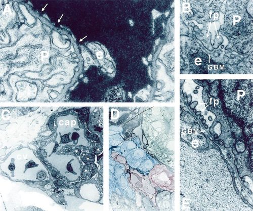
flh glomeruli contain appropriate glomerular cell types. In wildtype (A, B), podocytes (P) and endothelial cells (e) are found on opposite surfaces of the trilaminar glomerular basement membrane (GBM). Podocytes make well-formed, evenly spaced foot processes. Capillary endothelia show a fenestrated membrane morphology and pores are present (arrows in A). In flh glomeruli (C, D, E), blood-cell-containing capillaries (cap) are found adjacent to the cardinal vein (cv). In D, a vascular continuity (shaded in red) between the capillary space and the cardinal vein is visible and is surrounded by podocytes (shaded in blue). Capillary endothelia are intimately associated with morphologically identifiable podocytes in flh (asterisks in C). Podocytes and endothelia are organized on opposite sides of a well-formed and continuous GBM with well-formed lamina densa and lamina rarae (E). Podocytes appear morphologically normal with well-formed foot processes (fp). Significantly, endothelial cell membranes are nonfenestrated and continuous. Pores are absent. Occasionally, the membranes are detached from the GBM. Original magnifications: (A, B, E) 40,000x (C, D) 1600x PHENOTYPE:
|
Reprinted from Developmental Biology, 222(1), Majumdar, A. and Drummond, I.A., The zebrafish floating head mutant demonstrates podocytes play an important role in directing glomerular differentiation, 147-157, Copyright (2000) with permission from Elsevier. Full text @ Dev. Biol.