
- Title
-
The bHLH transcription factor Hand2 plays parallel roles in zebrafish heart and pectoral fin development
- Authors
- Yelon, D., Ticho, B., Halpern, M.E., Ruvinsky, I., Ho, R.K., Silver, L.M., and Stainier, D.Y.
- Source
- Full text @ Development
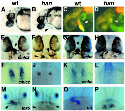
Myocardial defects in hans6 mutants. (A,C,E,G,I,K,M,O) Wild-type embryos; (B,D,F,H,J,L,N,P) hans6 mutant siblings. (A-D) Lateral views at 36 hpf, anterior to the left. (A,B) Bright-field images; mutants (B) display mild pericardial edema (arrowhead). (C,D) Immunofluorescent images of embryos stained with MF20 (TRITC) and S46 (FITC). In these double exposures, red fluorescence indicates MF20 staining of ventricular and somitic tissue, while yellow fluorescence indicates the overlap of S46 and MF20 staining in atrial tissue (Stainier and Fishman, 1992). (C) Wild-type embryos have a midline heart tube (arrowhead) with two distinct chambers, an anterior ventricle (red) and a posterior atrium (yellow). (D) hans6 mutants have two small clusters of myocardial tissue (arrowhead) that appear to be primarily atrial (yellow). (E-H, M-P) Dorsal views through the head at 33 hpf, anterior to the top. (M-P) are golden homozygotes. (I-L) Dorsal views of the myocardial precursors at the 15-somite stage (16.5 hpf). (E,F,I,J) In situ hybridization showing expression of the myocardial marker cmlc2 (Yelon et al., 1999). (E) Wildtype embryos express cmlc2 throughout the heart tube (arrowhead); (F) hans6 mutants have two small patches of cmlc2-expressing myocardial tissue (arrowheads). Younger wild-type embryos (I) have more myocardial precursors than hans6 mutant siblings (J). (G,H,K,L) Expression of the ventricular marker vmhc (Yelon et al., 1999). Wildtype embryos (G) express vmhc only within the future ventricle (arrowhead). hans6 mutants vary in amount of vmhc-expressing tissue: some hans6 mutants have no vmhc expression at this stage (data not shown), while others (H) have small populations of cells with weak vmhc expression (arrowheads). (K) Younger wild-type embryos express vmhc in a medial subset of myocardial precursors (Yelon et al., 1999); (L) vmhc expression is difficult to detect in hans6 mutants at this stage. (M,N) Expression of tbx5 in dorsal retina and heart tube (arrowhead) is apparent in wild-type embryos (M), but only dorsal retina expression is detectable in hans6 mutants (N, arrowheads indicate location of myocardium). (O,P) Expression of hrt in myocardium is apparent in wild-type embryos (O) and in hans6 mutants (P). EXPRESSION / LABELING:
PHENOTYPE:
|
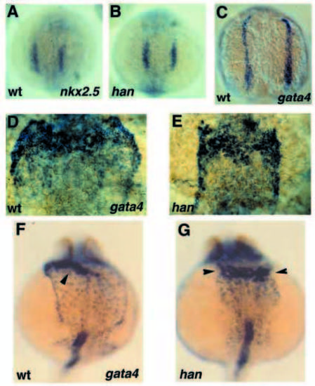
Normal precardiac mesoderm and aberrant LPM development in hans6 mutants. (A,B) Dorsal views at the 10-somite stage (14 hpf), anterior to the top. Wild-type (A) and hans6 mutant (B) siblings have indistinguishable nkx2.5 expression. (C-G) Dorsal views, anterior to the top, of gata4 expression at the 5-somite stage (approximately 12 hpf) (C), 15-somite stage (D,E) and 24 hpf (F,G). (C) Expression of gata4 in the anterior LPM begins in narrow bilateral stripes of cells that fuse medially by the 20-somite stage. (D) Before cardiac fusion begins, the gata4 expression domain has become much wider mediolaterally in wild-type embryos. (E) However, in hans6 mutants, gata4 expression remains as narrow bilateral stripes. (G) gata4 expression is maintained in the LPM and myocardia (arrowheads) of hans6 mutants. (F) The mutant LPM is narrow and dysmorphic in comparison to the broad sheet of gata4-expressing LPM in wild-type siblings (arrowhead indicates heart tube). We have obtained similar results with all other LPM markers, including gata5, gata6 and tbx5; that is, the expression domains of these genes expand in wild-type embryos but not in hans6 mutants. EXPRESSION / LABELING:
PHENOTYPE:
|

Pectoral fin defects in hans6 mutants. (A-D) Longitudinal sections through pectoral fin buds after in situ hybridization for tbx5 expression, anterior to the left. (A) At 32 hpf, a fin bud is forming in wild-type embryos. (B) hans6 mutants exhibit a delay in fin bud formation as well as a reduction in tbx5 expression at this stage. (C) In 48 hpf wild-type embryos, the pectoral fin is elongating and a chondrogenic condensation is forming. tbx5 expression is highest in the chondrogenic portion of the pectoral fin at this stage. (D) In hans6 mutants, a small undifferentiated fin bud expresses a reduced level of tbx5. (E) Dorsal views, anterior to the top, of a wild-type embryo (left) and a hans6 mutant (right) at 36 hpf. Embryos are golden homozygotes. shh expression is visible in the ZPA of each pectoral fin bud in wild-type embryos (arrowheads) but not in hans6 mutants. (F-I) Lateral views, anterior to the left, of pectoral fin buds from wild-type (F,H) and hans6 mutant (G,I) siblings at 32 hpf. (F) Wild-type embryos express hoxd-11 in a posterior portion (arrow) of the fin bud (outline indicated by arrowheads); (G) hans6 mutants never express hoxd-11 in the fin mesenchyme. (H) Wild-type embryos express hoxd-12 in a posterior portion (arrow) of the fin bud (outline indicated by arrowheads); (I) hans6 mutants never express hoxd-12 in the fin mesenchyme. (J,K) Dorsal views, anterior to the top, of the pectoral fin-forming region of the LPM in wild-type (J) and hans6 mutant (K) embryos at the 16-somite stage (17 hpf). The domain of tbx5 expression is expanded in wild-type embryos, but not in hans6 mutants. EXPRESSION / LABELING:
PHENOTYPE:
|

Comparison of wild-type, hans6, hanc99 and transheterozygote phenotypes. (A-O) Dorsal views, anterior to the top. (A,E,I,M,P) Wild-type embryos; (B,F,J,N,Q) hanc99 mutants; (C,G,K) transheterozygous mutants; (D,H,L,O,R) hans6 mutants. In all cases, the hans6 phenotype is most severe, the hanc99 phenotype is most mild and the transheterozygote phenotype is intermediate. (A-D) cmlc2 expression at 24 hpf; (E-H) tbx5 expression at 24 hpf. Arrowheads (F,G) indicate myocardial tissue. (I-L) gata4 expression at the 16-somite stage. (M-O) cmlc2 expression at the 16-somite stage. (P-R) Lateral views of pectoral fin buds, anterior to the left, demonstrating expression of dlx2 in the apical epidermal fold (arrowheads) at 48 hpf. Continuous and strong dlx2 expression is observed in the apical fold of wild-type pectoral fins (P) (Akimenko et al., 1994), but dlx2 maintenance is defective in hanc99 (Q) and hans6 (R) mutants. EXPRESSION / LABELING:
PHENOTYPE:
|

The s6 and c99 mutations disrupt hand2 genomic DNA. (A) Genomic structure of zebrafish hand2. The single intron is represented by a thick horizontal line; 5′ and 3′ UTRs are represented by thin rectangles and the coding regions of the two exons are represented by thick rectangles. A gray rectangle indicates the basic region and a black rectangle indicates the helix-loop-helix domain. (B) No fragment of the hand2 gene can be amplified by PCR from hans6 genomic DNA. Amplifications of a fragment from exon 1, a fragment from exon 2 and a control fragment from the end of a hand2-containing PAC are shown; alternating lanes represent reactions performed with wild-type and hans6 genomic DNA templates. (C) The hanc99 mutation is a ~5 kb insertion between bases 346 and 347 of the 5′ UTR. The insertion point is shown with a vertical line; the insertion is not drawn to scale. This insertion can lead to the missplicing of hand2 mRNA at cryptic splice sites at bases 233 and 578. The new ‘intron’ in this splice variant is represented by a thick line. Some 5′ UTR sequence and some coding sequence are spliced out in this variant. (D) hanc99 linkage testing was performed using a combination of three PCR primers (shown as red arrows in C, not drawn to scale). When the insertion is absent, the two primers flanking the insertion site amplify a small fragment, as in the case of a homozygous wild-type embryo (first lane). In the presence of the insertion, the flanking primers are ineffective using standard PCR conditions, but the 5′ primer and the primer complementary to the insertion can amplify a slightly larger fragment, as in homozygous hanc99 mutants (second-sixth lanes). (E-G) Dorsal views of cmlc2 expression in a wild-type embryo (E), a hans6 mutant sibling (F) and a hand2-injected hans6 mutant sibling (G). All panels are shown at the same magnification. Injection of hand2 mRNA can partially rescue the production of cmlc2-expressing myocardial precursors in hans6 mutants. Injection of hand2 mRNA does not seem to affect the production of myocardial precursors in wild-type embryos (data not shown). |

Expression pattern of zebrafish hand2. (A-F) In situ hybridization showing expression of hand2 in wild-type embryos. (A-C) Three views of the same embryo at tailbud stage (10 hpf), demonstrating hand2 expression in a continuous streak of the LPM. (A) Dorsal view of anterior part of the embryo, head at the top. (B) Dorsal view of posterior part of the embryo, tailbud at the bottom. (C) Lateral view, anterior to the left. (D,E) Two views of the same embryo at the 10-somite stage, demonstrating hand2 expression in a large portion of the LPM, with a gap (arrowheads in D) between the anterior and posterior expression domains. (D) Dorsal view of anterior part of the embryo, head at the top. (E) Dorsal view of posterior part of the embryo, tailbud at the bottom. (F) Dorsal view of the cardiogenic portion of the LPM, anterior at the top, demonstrating wider domains of hand2 expression at the 15-somite stage. (G,H) Dorsal views of the embryonic trunk at the 20-somite stage (19 hpf), anterior to the left, showing gene expression in the bilateral pectoral fin-forming regions of the LPM. hand2 expression (G) appears to overlap with a posterior portion of tbx5 expression (H). In H, intense expression of tbx5 in the dorsal retinae is visible through the yolk at the left side of the image. (I,J) Lateral views, anterior to the left, of pectoral fin buds at 32 hpf. Again, hand2 expression (I) appears to overlap with a posterior portion of tbx5 expression (J). (K) Dorsal view through the embryonic head at 28 hpf, anterior to the top. hand2 is expressed in the midline heart tube as well as within the bilateral sets of branchial arches. hand2 expression is also faintly visible in a broad sheet of LPM analogous to the gata4-expressing tissue shown in Fig. 2F. (L,M) Comparisons of hand2 expression in han mutant and wild-type embryos. Dorsal views, anterior to the top. (L) Wild-type (left) and hans6 mutant (right) embryos at the 5-somite stage. Wild-type embryos express hand2 in the LPM; hans6 mutant embryos do not express hand2 at this or any other stage. (M) hand2 expression at the 3-somite stage. hanc99 mutants (right) express lower levels of hand2 than wild-type siblings (left). hand2 expression levels are always reduced in hanc99 mutants, but the locations of hand2 expression are always normal. |