
- Title
-
Expression of sox11 gene duplicates in zebrafish suggests the reciprocal loss of ancestral gene expression patterns in development
- Authors
- De Martino, S., Yan, Y.-L., Jowett, T., Postlethwait, J.H., Varga, Z.M., Ashworth, A., and Austin, C.A.
- Source
- Full text @ Dev. Dyn.
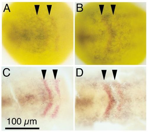
Expression of sox11a and sox11b during early development. A,B: Anterior dorsal views of whole-mount 1-somite stage embryos. C,D: Flat mounts of 1-somite stage embryos, labeled with mRNA probes for sox11a and sox11b(blue) and with krox20 (red). A-D: Anterior to the left. Arrow heads mark krox20 staining, representing prospective rhombomeres r3 and r5. Both sox11a and sox11b share an anterior medial domain of expression. In addition, sox11a is expressed as a broad stripe just anterior to, and overlapping with, the krox20 stripe in prospective rhombomere 3. In contrast, sox11b is expressed most highly between the krox20 stripes in prospective rhombomere 4, and weakly more posteriorly. Scale bar = 100 �m. EXPRESSION / LABELING:
|
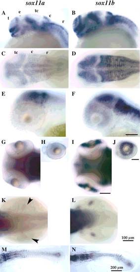
Sox11a and sox11b genes are expressed in overlapping and distinct regions of the embryo. A,B; E,F; H,J: Side views, anterior to the left. C,D; G,I; K-N: Dorsal views, anterior to the left. Scale bar = 100 �m unless otherwise indicated. t, telencephalon; e, epiphysis; tc, tectum; c, cerebellum; r, rhombencephalon. A,B: 24 hr embryos, sox11a is expressed in epiphysis (e), but not the tectum (tc); sox11b is expressed in the tectum, but not the epiphysis. C,D: 48 hr embryos, sox11b but not sox11a is expressed in the anterior lateral tectum at 48 hr. E,F: 48 hr embryos focused on the eye. G-J: 80 hr embryos, only low levels of sox11a are found in the eye at 80 hr, whereas sox11b is strongly expressed. K,L: 48 hr embryos, sox11b but not sox11a is expressed in the prospective fin buds at 48 hr. M,N: Flatmount of 14-somite stage embryos reveal differential expression of sox11a in the anterior somites and sox11b in the posterior somites. EXPRESSION / LABELING:
|
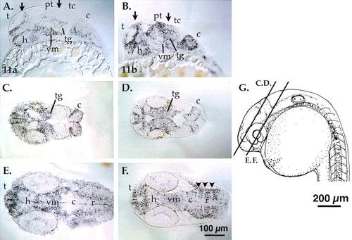
The sox11a and sox11b genes are expressed in overlapping and unique regions of the embryonic brain. A,B: Cells in the dorsal telencephalon, pretectum and tectum express sox11b (B), but not sox11a (A, arrows). Brain regions with similar sox11 gene expression domains include the telencephalon, diencephalon, ventral midbrain, and tegmentum. Ventral midbrain expression of sox11b is weaker than sox11a expression. The arrows indicate domains of differential gene expression in the dorsal telencephalon (left arrow in A and B) and the tectum (right arrows). C,D: sox11a and sox11b are expressed in the tegmentum and the cerebellum. The plane of sectioning is slightly more dorsal in C. E,F: In the ventral brain, ventral mesencephalic cells express sox11a and sox11b. However, sox11a expression is stronger posterior to the hypothalamus (posterior ventral diencephalon) and ventral midbrain than sox11b. Section 5F is a deep horizontal section, and median cells lacking sox expression in the cerebellum are in the floor plate region. A,B sagittal sections; C-F horizontal sections, as indicated in G. t, telencephalon; h, hypothalamus; tg, tegmentum; pt, pretectum; tc, tectum; vm, ventral mesencephalon; c, cerebellum; r, rhombencephalon. Scale bars = 100 �m in A-F, 200 �m in G. EXPRESSION / LABELING:
|

Sox11 gene expression in somites of zebrafish and quail. A,B: Flatmounts of 14-somite stage zebrafish embryos, revealing differential expression of sox11a in the nine anterior somites and sox11b in the five posterior somites (black arrows). C: A wholemount of a stage 13 quail embryo showing expression of SOX11 in all the somites. Scale bars = 200 �m and 1mm. |

Unillustrated author statements EXPRESSION / LABELING:
|