
Image
|
Figure Caption
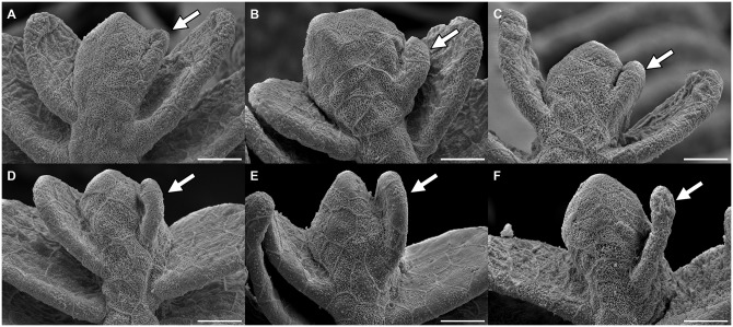
Fig 6
Read from A to F: the images show the sprouting stages of a new filament (arrow) on the right of the primary filament tip with initial thickening, progressive separation from the tip and growth of the newly developed filament. Scale bar: 15
Figure Data
Acknowledgments
This image is the copyrighted work of the attributed author or publisher, and
ZFIN has permission only to display this image to its users.
Additional permissions should be obtained from the applicable author or publisher of the image.
Full text @ PLoS One