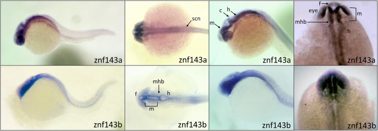
IMAGE

Fig. 3

ID
ZDB-IMAGE-200130-9
Genes
Source
Figures for Huning et al., 2020
Image
Figure Caption

Fig. 3

Similar spatial expression of znf143a and znf143b in 24hpf embryos. Antisense digoxigenin riboprobes against the znf143a and znf143b were used for whole-mount in situ hybridization assays. The probe for znf143b targeted the last exon of the coding region (approximately 186 nt corresponding to the last 61 amino acids) and the 3’UTR of the gene, while the probe for znf143a was designed solely against the 3’UTR. Embryos used were fixed and stained at 24hpf. The panels illustrate views from four different embryos for each probe, with the right-most panels showing an enlarged dorsal view of the head. We note that in all experiments, the znf143b probe produced fainter staining, most likely because of a smaller than optimal riboprobe that was necessary to ensure gene specificity. Specific head, brain and neural structures are identified with the following abbreviations: f, forebrain; m, midbrain; h, hindbrain; mhb, midbrain hindbrain boundary; c, cerebellum; scn, spinal cord neurons

Figure Data
Acknowledgments
This image is the copyrighted work of the attributed author or publisher, and ZFIN has permission only to display this image to its users. Additional permissions should be obtained from the applicable author or publisher of the image. Full text @ BMC Mol Cell Biol